The Skeletal System Essay
Introduction, axial portion of the skeleton, appendicular portion of the skeleton, functions of the skeleton, relationship between the skeletal system and the muscular system, sexual differences in skeletons, clinical conditions and disorders that affect the skeleton, works cited.
Movement is vital for all of you because it provides you with the opportunity to live your lives to the full. Just as other human beings, you fall and stand up to continue moving forward. But what provides you with this opportunity? It is your skeletal system. It does not only facilitate your physical activity but also supports and protects your bodies. This system consists of hundreds of bones that are full of calcium, which makes them strong enough to carry your weight. Bones are connected with the help of joints that facilitate motion. The majority of you were born with about 300 bones that fuse with the course of time so that now you have only 206 bones. They all are divided into two parts: axial and appendicular skeletons.
Your axial portion of skeleton is composed of “the skull, the vertebral column, and the thoracic cage” ( Skeletal System: Bones and Joints 120). Due to its location, it manages to protect your brain and spinal cord from injuries. In addition to that, it supports the organs in the ventral body cavity so that you do not need to carry them in your hands.
Twenty-two bones that are separated into two parts form the skull. You have 8 bones of the cranial cavity that are known as braincase. They surround your brain so that you do not hurt it when fall or receive a headnut. The rest of the bones (there are 14 of them) form your face. They are tightly connected to one another so that your nose is always in the right place. The only exception is the mandible that makes chewing possible. Otherwise, how would you eat? Minimal movement can also be observed within the middle ears. Each of them includes 3 auditory ossicles that are hidden deep in your head.
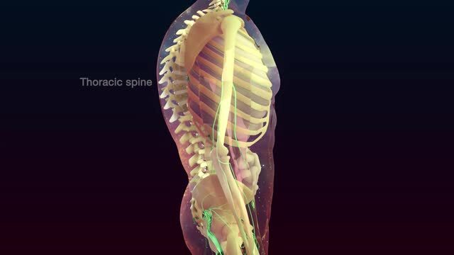
The vertebral column, or backbone, usually consists of “7 cervical vertebrae, 12 thoracic vertebrae, 5 lumbar vertebrae, 1 sacral bone, and 1 coccyx bone” ( Skeletal System: Bones and Joints 125). It is the central axis of the skeleton that has four major curvatures. Normally, the cervical and the lumbar regions curve anteriorly. The thoracic, as well as the sacral and coccygeal regions, curves posteriorly. However, considering the way you sit, abnormal curvatures are widespread.
The thoracic or the rib cage protects your organs and supports them. All in all, human beings have 24 ribs that are divided into 12 pairs, but you can recount them to make sure. They are categorized according to their attachment to the sternum. Thus, a direct attachment by costal cartilages is true (1-7); an attachment by a common cartilage is false (8-12); and the absence of attachment resorts to floating ribs (11-12). The sternum, or breastbone, consists of three parts: “the manubrium, the body, and the xiphoid process” ( Skeletal System: Bones and Joints 129).
Your appendicular skeleton consists of the bones of limbs and girdles so that you have:
- “4 bones in the shoulder girdle (clavicle and scapula each side).
- 6 bones in the arm and forearm (humerus, ulna, and radius).
- 58 bones in the hands (carpals 16, metacarpals 10, phalanges 28, and sesamoid 4).
- 2 pelvis bones.
- 8 bones in the legs (femur, tibia, patella, and fibula).
- 56 bones in the feet (tarsals, metatarsals, phalanges, and sesamoid)” (“The Axial & Appendicular Skeleton” par. 4).
What would you be without this part of skeleton? Imagine that it is a big 3D puzzle, gathering all these bones together in a right order, you will build your arms and legs with all details. These are all movable parts that allow you to run, dance, write, and even hug your nearest and dearest. Even though the axial skeleton seems to be more important because it is connected with your brain, the appendicular portion of the skeleton contains about 60% of all your bones, which means that its importance should not be undervalued.
As you have already understood, your skeleton maintains a lot of different functions. Some of them, such as movement and support, were already mentioned. But let us discuss them all in detail.
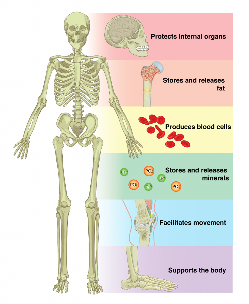
- Support. Your bodies are supported by the skeleton so that you can change your position to vertical one and stand strait. Without it, you would be able only to lie because of the gravitation. This function is provided by many bones but the long ones seem to be the leaders in this competition. For instance, those that are in legs, support the trunk. Similarly, vertebras support one another so that eventually the firs one provides support to the skull. In addition to that, they support the organs and ensure that they do not change their positions.
- Protection. The skeleton also protects you. For example, the skull prevents fatal brain injuries. The rib cage protects such vital organs as the heart and lungs. It also takes care of your abdominal organs ensuring that they develop normally.
- Movement. The function of bodily motion allowed you to come here today. However, it is critical to remember that it is maintained not only due to the bones but also with the help of the muscular system.
- Mineral and energy storage. From the outer side of your bones, there is a tissue that serves as a storage. It gathers calcium and phosphorus and withdraws them to maintain appropriate blood levels. In addition to that, mature bones store yellow marrow. It consists of fat almost totally and provides you with energy for various activities.
- Blood-cell formation. The inner core of your bones takes part in the formation of blood cell and platelet. It is known as bone marrow or red marrow. Platelet is vital for you because it ensures your ability to heal wounds while blood cells spread oxygen and destroy infectious cells (CAERT 3).
Have you ever thought of the way our movement are maintained? Even a simple nod of the head requires the cooperation between the skeletal and muscular systems. Muscles ensure movement of our body through the attachment to the bones. All in all, there are about 700 of them, which is an enormous amount that comprises about 50% of your weight.
So what happens in your body when you moves? When you want to move, your brain sends a message for the body to release energy. In medical terms, it is called adenosine triphosphate. Affecting your muscles, it makes them contract or shorten. Shortened muscles pulls bones at their insertion point. Thus, the angle between the bones connected by a joint shortens. Relaxation is maintained when the opposing muscle extends and pulls a bone to its initial position.
Human skeletons seem to be similar, as they contain the same bones. However, you should remember that their characteristics differ depending on the gender. For example, women have lighter pelvis bones that form a shorter cavity with less dimensions. It has less prominent marking for muscles and more circular pelvic brim. The sacral bones of men are longer and narrower, which makes them more massive. Their femur is also longer and heavier. Its texture is rough unlike women’s smooth.
Muscle marking is more developed and shaft is less oblique. The head of men’s femur is larger and trochanters are more prominent. The femoral neck angle in males is more than 125 and in females is less than 125. Women’s sternum is less than twice the length of manubrium and larger in men. Differences in skull include greater capacity, thicker walls, more marked muscular ridges, prominent air sinuses, smoother upper margin of orbit, less vertical forehead, and heavier cheekbones in males.
Hopefully, it will never affect any of you but the skeleton may be affected by tumours that cause bone defects. People may have skeletal developmental disorders including gigantism, dwarfism, osteogenesis imperfecta, and rickets lead to abnormal body sizes, brittle bones, and growth retardation. Bacterial infections cause inflammation and lead to bone destruction.
Decalcification, including the known to you osteoporosis, reduces bone tissue and softens bones. Joint disorders often deal with inflammation. For instance, arthritis. They are often influenced by age and physical activity. In this way, degradation of joints is observed in the elderly but can be delayed due to regular exercises. The abnormal curvatures of the spine may also cause health issues. That is why you should pay attention to your back posture and avoid kyphosis (a hunchback condition), lordosis (a swayback condition), and scoliosis (an abnormal lateral curvature).
CAERT. Structures and Functions of the Skeletal System . 2014. Web.
Skeletal System: Bones and Joints. 2012. Web.
“ The Axial & Appendicular Skeleton. ” TeachPE , 2017. Web.
- Chicago (A-D)
- Chicago (N-B)
IvyPanda. (2023, December 13). The Skeletal System. https://ivypanda.com/essays/the-skeletal-system/
"The Skeletal System." IvyPanda , 13 Dec. 2023, ivypanda.com/essays/the-skeletal-system/.
IvyPanda . (2023) 'The Skeletal System'. 13 December.
IvyPanda . 2023. "The Skeletal System." December 13, 2023. https://ivypanda.com/essays/the-skeletal-system/.
1. IvyPanda . "The Skeletal System." December 13, 2023. https://ivypanda.com/essays/the-skeletal-system/.
Bibliography
IvyPanda . "The Skeletal System." December 13, 2023. https://ivypanda.com/essays/the-skeletal-system/.
- Articular and Muscular Systems
- The Muscular System of a Human Body
- Aspects of the Skeletal System
- Neuropsychological Tests Reliability Following Concussion
- Physicians, Their Roles and Responsibilities
- Prevalence of Sleep Disorders among Medical Students
- Human Physical Performance Under Adverse Conditions
- Tongue and Why It Is Unique

- school Campus Bookshelves
- menu_book Bookshelves
- perm_media Learning Objects
- login Login
- how_to_reg Request Instructor Account
- hub Instructor Commons
- Download Page (PDF)
- Download Full Book (PDF)
- Periodic Table
- Physics Constants
- Scientific Calculator
- Reference & Cite
- Tools expand_more
- Readability
selected template will load here
This action is not available.

14: Skeletal System
- Last updated
- Save as PDF
- Page ID 92678

- Tara Jo Holmberg
- Northwestern Connecticut Community College
This chapter describes the structure and functions of the skeletal system and its two major divisions, the axial skeleton and the appendicular skeleton. It details the structure of bone, how bones grow, and how they are remodeled and repaired. The chapter also explains how joints work and how they are classified as well as the causes and effects of major skeletal system disorders.
- 14.1: Case Study: Your Support System Amari loves wearing high heels when they go out at night, especially stilettos. They know high heels are not the most practical shoes, but Amari likes how they look. Lately Amari has been experiencing pain in the balls of thier feet—the area just behind the toes. Even when they trades heels for comfortable sneakers, it still hurts when Amari stands or walks.
- 14.2: Introduction to the Skeletal System The skull and cross-bones symbol has been used for a very long time to represent death, perhaps because after death and decomposition, bones are all that remain. Many people think of bones as being dead, dry, and brittle. These adjectives may correctly describe the bones of a preserved skeleton, but the bones of a living human being are very much alive. Living bones are also strong and flexible. Bones are the major organs of the skeletal system.
- 14.3: Divisions of the Skeletal System This somewhat macabre display can be viewed at the Slovak National Museum in Bratislava, Slovakia. The skulls are meant to represent normal human skeletal anatomy. The skull is part of the axial skeleton, which is one of the two major divisions of the human skeleton. The other division is the appendicular skeleton.
- 14.4: Structure of Bone Do you recognize the food item in the top left of this photo? It's roasted bone marrow, still inside the bones. It's considered a delicacy in some cuisines. Marrow is a type of tissue found inside many animal bones, including our own. It's a soft tissue that in adults may be mostly fat. You'll learn more about bone marrow and other tissues that make up bones when you read this concept.
- 14.5: Bone Growth, Remodeling, and Repair Did you ever break a leg or other bone, like the man looking longingly at the water in this swimming pool? Having a broken bone can really restrict your activity. Bones are very hard, but they will break, or fracture, if enough force is applied to them. Fortunately, bones are highly active organs that can repair themselves if they break. Bones can also remodel themselves and grow. You'll learn how bones can do all of these things in this concept.
- 14.6: Joints Joints are locations at which bones of the skeleton connect with one another. A joint is also called an articulation. The majority of joints are structured in such a way that they allow movement. However, not all joints allow movement. Of joints that do allow movement, the extent and direction of the movements they allow also vary.
- 14.7: Disorders of the Skeletal System The woman on the right in this image has a deformity in her back commonly called dowager's (widow's) hump, because it occurs most often in elderly women. Its medical name is kyphosis, and it is defined as excessive curvature of the spinal column in the thoracic region. The curvature generally results from fractures of thoracic vertebrae. As the inset drawings suggest, these fractures may occur due to a significant decrease in bone mass, which is called osteoporosis. Osteoporosis is one of the mo
- 14.8: Case Study Conclusion: Heels and Chapter Summary You may have seen signs indicating that high-heeled shoes are not allowed on certain walking surfaces because of the risk of injury. High heels affect a person's balance, and wearers can easily twist their ankle on uneven or slippery surfaces, causing a sprain or even a fracture. Besides twisting an ankle, wearing high heels on a regular basis can cause a variety of other negative health consequences—some of which may be long-lasting.

- school Campus Bookshelves
- menu_book Bookshelves
- perm_media Learning Objects
- login Login
- how_to_reg Request Instructor Account
- hub Instructor Commons
- Download Page (PDF)
- Download Full Book (PDF)
- Periodic Table
- Physics Constants
- Scientific Calculator
- Reference & Cite
- Tools expand_more
- Readability
selected template will load here
This action is not available.
5.1: Introduction to Bone Tissue and the Skeletal System
- Last updated
- Save as PDF
- Page ID 22276
- Whitney Menefee, Julie Jenks, Chiara Mazzasette, & Kim-Leiloni Nguyen
- Reedley College, Butte College, Pasadena City College, & Mt. San Antonio College via ASCCC Open Educational Resources Initiative
Chapter Learning Objectives
After studying this chapter, you will be able to:
- List and describe the functions of bones
- Describe the classes of bones
- Discuss the process of bone formation and development
- Explain how bone repairs itself after a fracture
- Discuss the effects of exercise, nutrition, and hormones on bone tissues
Bones make good fossils. While the soft tissue of a once living organism will decay and fall away over time, bone tissue will, under the right conditions, undergo a process of mineralization, effectively turning the bone to stone. A well-preserved fossil skeleton can give us a good sense of the size and shape of an organism, just as your skeleton helps to define your size and shape. Unlike a fossil skeleton, however, your skeleton is a structure of living tissue that grows, repairs, and renews itself. The bones within it are dynamic and complex organs that serve a number of important functions, including some necessary to maintain homeostasis.

Contributors and Attributions
OpenStax Anatomy & Physiology (CC BY 4.0). Access for free at https://openstax.org/books/anatomy-and-physiology
Chapter 7- The Skeletal System
Introduction to the skeletal system, learning objectives.
- Describe the functions of the skeletal system.
- Distinguish between long bones, short bones, flat bones, and irregular bones and provide an example of each.
- Identify the anatomical features of a long bone
- Describe the microscopic structure of compact bone, and compare it with that of spongy bone.
- Identify all the bones of the axial and appendicular skeleton.
- Describe various skeletal joints and the movements possible
- Describe the steps involved in bone development and bone repair
- Describe the effect exercise has on bone tissue
- Describe the disorders of the skeletal system
- Describe the effects of hormones on bone tissue, the process of calcium homeostasis
Bone , or osseous tissue , is a hard, dense connective tissue that forms most of the adult skeleton, the support structure of the body. In the areas of the skeleton where bones move (for example, the ribcage and joints), cartilage , a semi-rigid form of connective tissue, provides flexibility and smooth surfaces for movement.
The skeletal system is the body system composed of bones and cartilage and performs the following critical functions for the human body:
- protection of vital structures, such as the spinal cord, brain, heart, and lungs.
- support of body structures.
- body locomotion through coordination with the muscular system.
- hematopoiesis, or generation of blood cells, within the red marrow spaces of bones.
- storage and release of the inorganic minerals calcium and phosphorous, which are needed for functions such as muscle contraction and neural signal conduction.
The most apparent functions of the skeletal system are the gross functions—those visible by observation. Simply by looking at a person, you can see how the bones support, facilitate movement, and protect the human body.

Figure 1. Bones Protect Brain. The cranium completely surrounds and protects the brain from non-traumatic injury.
Just as the steel beams of a building provide a scaffold to support its weight, the bones and cartilage of your skeletal system compose the scaffold that supports the rest of your body. Without the skeletal system, you would be a limp mass of organs, muscle, and skin.
Bones also facilitate movement by serving as points of attachment for your muscles. While some bones only serve as a support for the muscles, others also transmit the forces produced when your muscles contract. From a mechanical point of view, bones act as levers and joints serve as fulcrums.
Unless a muscle spans a joint and contracts, a bone is not going to move. For information on the interaction of the skeletal and muscular systems, that is, the musculoskeletal system, seek additional content.
Bones also protect internal organs from injury by covering or surrounding them. For example, your ribs protect your lungs and heart, the bones of your vertebral column (spine) protect your spinal cord, and the bones of your cranium (skull) protect your brain (Figure 1).
Career Connection: Orthopedist
An orthopedist is a doctor who specializes in diagnosing and treating disorders and injuries related to the musculoskeletal system. Some orthopedic problems can be treated with medications, exercises, braces, and other devices, but others may be best treated with surgery (Figure 2).

Figure 2. Arm Brace. An orthopedist will sometimes prescribe the use of a brace that reinforces the underlying bone structure it is being used to support. (credit: Juhan Sonin)
While the origin of the word “orthopedics” (ortho- = “straight”; paed- = “child”), literally means “straightening of the child,” orthopedists can have patients who range from pediatric to geriatric. In recent years, orthopedists have even performed prenatal surgery to correct spina bifida, a congenital defect in which the neural canal in the spine of the fetus fails to close completely during embryologic development.
Orthopedists commonly treat bone and joint injuries but they also treat other bone conditions including curvature of the spine. Lateral curvatures (scoliosis) can be severe enough to slip under the shoulder blade (scapula) forcing it up as a hump. Spinal curvatures can also be excessive dorsoventrally (kyphosis) causing a hunch back and thoracic compression. These curvatures often appear in preteens as the result of poor posture, abnormal growth, or indeterminate causes. Mostly, they are readily treated by orthopedists. As people age, accumulated spinal column injuries and diseases like osteoporosis can also lead to curvatures of the spine, hence the stooping you sometimes see in the elderly.
Some orthopedists sub-specialize in sports medicine, which addresses both simple injuries, such as a sprained ankle, and complex injuries, such as a torn rotator cuff in the shoulder. Treatment can range from exercise to surgery.
Mineral Storage, Energy Storage, and Hematopoiesis

Figure 3. Head of Femur Showing Red and Yellow Marrow. The head of the femur contains both yellow and red marrow. Yellow marrow stores fat. Red marrow is responsible for hematopoiesis. (credit: modification of work by “stevenfruitsmaak”/Wikimedia Commons)
On a metabolic level, bone tissue performs several critical functions. For one, the bone matrix acts as a reservoir for a number of minerals important to the functioning of the body, especially calcium, and potassium. These minerals, incorporated into bone tissue, can be released back into the bloodstream to maintain levels needed to support physiological processes. Calcium ions, for example, are essential for muscle contractions and controlling the flow of other ions involved in the transmission of nerve impulses.
Bone also serves as a site for fat storage and blood cell production. The softer connective tissue that fills the interior of most bone is referred to as bone marrow (Figure 3). There are two types of bone marrow: yellow marrow and red marrow. Yellow marrow contains adipose tissue; the triglycerides stored in the adipocytes of the tissue can serve as a source of energy. Red marrow is where hematopoiesis —the production of blood cells—takes place. Red blood cells, white blood cells, and platelets are all produced in the red marrow.
Classify bones according to their shapes
The 206 bones that compose the adult skeleton are divided into five categories based on their shapes (Figure 4). Their shapes and their functions are related such that each categorical shape of bone has a distinct function.

Figure 4. Classifications of Bones. Bones are classified according to their shape.
Long Bones: A long bone is one that is cylindrical in shape, being longer than it is wide. Keep in mind, however, that the term describes the shape of a bone, not its size. Long bones are found in the arms (humerus, ulna, radius) and legs (femur, tibia, fibula), as well as in the fingers (metacarpals, phalanges) and toes (metatarsals, phalanges). Long bones function as levers; they move when muscles contract.
Short Bones: A short bone is one that is cube-like in shape, being approximately equal in length, width, and thickness. The only short bones in the human skeleton are in the carpals of the wrists and the tarsals of the ankles. Short bones provide stability and support as well as some limited motion.
Flat Bones: The term flat bone is somewhat of a misnomer because, although a flat bone is typically thin, it is also often curved. Examples include the cranial (skull) bones, the scapulae (shoulder blades), the sternum (breastbone), and the ribs. Flat bones serve as points of attachment for muscles and often protect internal organs.
Irregular Bones: An irregular bone is one that does not have any easily characterized shape and therefore does not fit any other classification. These bones tend to have more complex shapes, like the vertebrae that support the spinal cord and protect it from compressive forces. Many facial bones, particularly the ones containing sinuses, are classified as irregular bones.
Sesamoid Bones: A sesamoid bone is a small, round bone that, as the name suggests, is shaped like a sesame seed. These bones form in tendons (the sheaths of tissue that connect bones to muscles) where a great deal of pressure is generated in a joint. The sesamoid bones protect tendons by helping them overcome compressive forces. Sesamoid bones vary in number and placement from person to person but are typically found in tendons associated with the feet, hands, and knees. The patellae (singular = patella) are the only sesamoid bones found in common with every person. Table 1 reviews bone classifications with their associated features, functions, and examples.
- Chapter 6. Authored by : OpenStax College. Provided by : Rice University. Located at : http://cnx.org/contents/[email protected]. . Project : Anatomy & Physiology. License : CC BY: Attribution . License Terms : Download for free at http://cnx.org/content/col11496/latest/
- Essay Editor
The Skeletal System
1. introduction.
The skeletal system consists of bones and their associated connective tissues - including cartilage, tendons, and ligaments. It serves the following functions: • Support: The bones of the legs act as pillars to support the body trunk when standing, and the ribcage supports the thoracic wall. • Protection: Bones surround and protect soft and vital organs. For example, the skull protects the brain, the vertebrae shield the spinal cord, and the ribcage protects the heart and lungs. • Movement: Bones serve as points of attachment for most skeletal muscles. • Mineral and growth factor storage: Bone is a reservoir for minerals, most importantly calcium and phosphorus. When required, these minerals can be released into the bloodstream to serve other critical functions. • Blood cell formation: Hematopoiesis occurs within the marrow cavities of certain bones. • Triglyceride (fat) storage: Yellow marrow functions as a storage area for triglycerides. Due to these important functions, the study of the skeletal system is critical to understanding human anatomy and thus the medical and health sciences. It provides a basic framework for our body, and without it, our soft organs and tissues would have no protection or anchor. Understanding these functions can provide insight into numerous malfunctions and help diagnose and treat various medical conditions.
1.1. Definition and Importance
The adult human skeletal system consists of 206 bones and several different types of tissues. It is divided into two functional parts. The axial skeleton consists of 80 bones. It includes the skull, the ossicles of the ear, the hyoid bone, the vertebral column, and the thoracic cage. The appendicular skeleton, which is attached to the axial skeleton, consists of 126 bones. It includes the pectoral girdles, the upper limbs, the pelvic girdle, and the lower limbs. The varied shapes and sizes of the bones that constitute the human skeleton reflect a number of different evolutionary adaptations. Although the most conspicuous role of the skeleton is to provide a solid framework for the body and to protect the internal organs, it also serves as a system of levers that the muscles use to move the body. In addition, bone is a reservoir for minerals such as calcium and phosphate, and it is the production site for blood cells and white blood cells.
1.2. Components of the Skeletal System
The skeletal system has several functions, the most obvious of which is to provide a framework for the body. Other functions are to act as a protective case for the internal organs and to provide a system of levers which the muscular system uses to move the body. Though it consists of all the bones in the body, the human skeletal system is not a static organ. Blood and stem cells in the bone marrow are tissue from which the rest of the body can be grown and repaired. The human skeletal system is divided into the axial skeleton and the appendicular skeleton. The axial skeleton includes the skull, spinal column, ribs, and breastbone. The skull is further divided into the cranium which houses the brain and the facial bones. The spinal column is comprised of 33 interlocking vertebrae. The appendicular skeleton is made up of the bones of the upper and lower limbs and the girdles which connect the limbs to the axial skeleton. A long bone is one that is cylindrical and elongated and has growth plates at the end. All the limb bones are long bones. A short bone is one that is cube-like in shape and is approximately equal in length, width, and thickness. The wrist is comprised of eight short bones arranged in two rows. The patella is also classified as a short bone. A flat bone is usually thin and curved. They serve as protective plates for internal organs and also have a large surface area for muscle attachment. The pelvis, sternum, and cranium are all flat bones. An irregular bone is one that does not fit into any of the previous categories. This is because it has a complex shape with several points of attachment. The vertebrae is an irregular bone.
2. Functions of the Skeletal System
The skeleton has five main functions, which are divided into minor categories. The first main function of the skeleton is to provide support for the body. It does this by acting as a hard framework, which supports all the soft tissues and provides a base for movement. Bone is very lightweight but still very strong, making it perfect for its supportive role. The second function of the skeleton is protection of the internal organs from external forces. An example of this would be the ribcage, which protects the heart and lungs, or the skull, which surrounds and protects the brain. Joints are the locations where bones meet. They make possible any movement between the bones. Most joints allow for movement between the bones they connect, although some joints do not allow any movement at all. The movement of bones at a joint is brought about by the contraction of muscles, which are attached to the bones by tendons. The shape of a bone often determines the type of movement possible. For example, a ball and socket joint at the hip allows a wide range of movement, but a pivot joint at the neck only allows rotation. The third function of the skeleton is movement. By using its many muscles, the body is able to walk, run, and move about. Muscles are able to pull on bones to make them move at special structures called joints. At the point where the muscle is attached to a bone, there is normally a bulge. This is because the bone is actually a bit thicker there and is called a process (it is also the place from which the bone gets its name). When a muscle pulls on a bone, it must overcome the resistance provided by the weight of the body and that of anything being carried. Because of this, many bones have special ridges or bumps which function to anchor the tendons of important muscles. An example of this would be the knees formed by the flaring of the femur and tibia or the knuckles at the hand. The fourth function of the skeleton is the storage of mineral reserves. Some minerals are needed to perform vital metabolic functions. When these minerals are not present in sufficient amounts in the blood, the body will take them from the bones. This most commonly involves calcium and phosphorus, which are stored in crystals in the structure of the bones. Calcium reserves in the bones can be mobilized to maintain a constant level of calcium in the blood, which is necessary to enable normal muscle and nerve function. In situations where too much calcium is taken from the bones (for example, through not eating enough calcium-containing foods), it can result in a condition known as osteoporosis, in which the bones become weak and more likely to break.
2.1. Support and Protection
Just holding the body up and keeping it stable, it helps in securing the soft organs so that fatal injuries do not occur. For example, the ribcage protects the heart and lungs, which are the most delicate organs. The cranium is almost like a protective helmet, which is imperative as the brain is such a delicate organ. The vertebrae also help to keep the spinal cord safe. It also serves for muscular attachment so that movement is possible. The skeletal system is a system made up of 206 bones, as well as a few teeth, and is rigid and sturdy. Its main functions are to support, provide a framework for the attachment of muscles and other tissues, support soft organs, and maintain the body's shape. The second function is protection. This is where the skeletal system comes into its name. This occurs by providing a framework for the attachment of muscles and other tissues, supporting soft organs, and maintaining the body's shape. Now, since the shoe incident, your feet have been greatly misshaped. Don't worry! The last function of the skeletal system is lifelong. It provides a place for mineral storage, especially calcium and phosphorus, which gives bones their hardness and rigidity. This occurs by the instruction of bone to release small amounts of minerals into the bloodstream for distribution to the body when minerals are low in the body. If the minerals in bones are excessively low, the body will instruct the bones to retrieve minerals from the bones to restore them to an appropriate level in the blood. Another important function for older folks is that bones produce growth factors for other tissues, and one important growth factor is blood cell formation. The final function of the skeletal system is to act as a hormone producer. An example of a detrimental hormone in action is the sex hormones, which are secreted by the pelvis. This can lead to bone weakening or joint loosening in the area. A good function of a bone hormone is when there is an excessive decrease in calcium level hormone, it increases the rate of calcium absorption in the gut, which will restore calcium to an appropriate level in the body.
2.2. Movement and Locomotion
In order to understand the role of the skeletal system in movement, we must return to the lever system. The muscles provide the effort; the joints act as the fulcrum; the bones provide the lever; and the resistance is whatever is being moved. In this model, bones serve to transmit the forces applied by muscles which move them in order to generate useful movement at the joints. Joints close to the body, such as the hip and shoulder, are acted upon by the weight of the body which creates a moment or rotational force about the joint which must be balanced by an equal force acting in the opposite direction. A good example of this is the almighty struggle that takes place in the first few seconds of a tug-of-war match, with the force exerted by each team being translated through the legs into an attempt to move the hip joint. By taking a look at the relative size and orientation of the bony markers on a humerus it is possible to deduce the strength of the muscles acting across a joint and the resulting capabilities of that joint. The shape of a bone and the nature of its articulation determine the movement possible at a joint. For example, the ball and socket joints such as the hip allow movement in all planes and are capable of angular and rotational movement. These joints are well served by the rounded and spherical head of the humerus or femur which provides maximum support and strength at the expense of material; the force exerted by weight or muscle can be considerable and the load on the joint can be very high. The structural design of long bones allows the force exerted on them to be safely transmitted across the length of the bone to a joint where movement will take place. This is achieved by having a large body which is tapered at the ends to produce a neck which is the site of articulation, an excellent example being the femur.
2.3. Blood Cell Production
Hematopoiesis, or the process of blood cell production, occurs in the marrow of long bones in the body. Blood cells originate from the same type of unspecialized cell, called the haemocytoblast, which is derived from the mesenchyme. These unspecialized cells are stimulated to multiply and differentiate into specialized cells by the hormone erythropoietin. There are a few forms of marrow in the body - red and yellow marrow. Red marrow is responsible for hematopoiesis and is located in the spongy bone. Yellow marrow is mainly a fat storage area and can be converted to red marrow if the body is severely anemic. All red marrow has the ability to produce more red blood cells. In infants, blood cells are produced in the medullary cavity of the spongy bone. As the child grows, the site of hematopoiesis moves towards the epiphyses of the long bones. This area remains the primary site of blood cell production in adults. With increasing age, hematopoiesis in the axial skeleton and proximal epiphyses decreases, and in old age, it ceases altogether. At this stage, it goes beyond the scope of normal skeletal function.
3. Common Skeletal Disorders
Osteoporosis is a condition that causes bones to become less dense and more fragile. Although bone density naturally decreases with age, osteoporosis speeds up this process, increasing the risk of breaking a bone. The word "osteoporosis" literally means "porous bone." When viewed under a microscope, healthy bone looks like a honeycomb. When osteoporosis occurs, the holes and spaces in the honeycomb are much larger than in healthy bone. Osteoporotic bones have lost density or mass and contain abnormal tissue structure. As bones become less dense, they weaken and are more likely to break. If you're 50 or older and have broken a bone, ask your doctor or healthcare provider about a bone density test. A bone density test measures the strength and density of your bones by using x-rays. The most accurate test is a dual energy x-ray absorptiometry, or DXA test. The best test to predict your risk of fractures uses the results of the DXA test and other factors that you give your healthcare provider. This is called a Fracture Risk Assessment. Osteoporosis is a major public health threat for an estimated 44 million Americans, or 55 percent of the people 50 years of age and older. In the United States, 10 million individuals are estimated to already have the disease and almost 34 million more are estimated to have low bone mass, placing them at increased risk for osteoporosis. It is often thought of as an older person's disease, but it can strike at any age. Arthritis is a disease that affects many Americans as they grow older. It is usually associated with pain and/or swelling in the joints. There are over 100 different types of arthritis, many of which are often chronic and can have severe effects on an individual's quality of life. Some types of arthritis can also affect other tissues and organs including the skin and immune system. One of the most common types of arthritis is called osteoarthritis. Osteoarthritis is a degenerative joint disease, which affects the articular cartilage and the bones. It is often the result of years of wear and tear on the joints, and is usually associated with joint pain and stiffness. Another type of arthritis that affects the articular surface is rheumatoid arthritis. Rheumatoid arthritis is a systemic disease, often affecting extra-articular tissues throughout the body. The most common feature of rheumatoid arthritis is persistent inflammation of synovium, which leads to joint damage. Joint involvement is often symmetrical, meaning that if one joint is affected the same joint on the opposite side of the body is usually affected as well. Scoliosis is a lateral (toward the side) curvature in the normally straight vertical line of the spine. The normal spine looks straight when viewed from the front, but has three gentle curves when seen from the side. These are known as the curves of the spine, and they are necessary in order to help the spine withstand stress from body weight and gravity. A healthy spine would look like an "S" from the side. A person with scoliosis will have a spine that looks more like a "C" from the back, because the spine has laterally curved, often in more than one place. Scoliosis is a common disease, affecting around 3% of the population. There are many different causes for scoliosis, but the most common is a condition called idiopathic scoliosis. This condition occurs in children between the age of 10-13 years, and affects 2% of this population. An abnormal gene that is thought to be inherited from the child's parents causes the development of scoliosis. This gene does not directly cause scoliosis, but alters the normal pattern of growth of the child's spine which results in the development of scoliosis.
3.1. Osteoporosis
Globally, it is the second most common disease after cardiovascular disease. Osteoporosis is defined in women as a decrease of 35% of their bone density by the time they reach the age of 60. This means that the bone has become weak and brittle and likely to fracture through a minor injury that in normal circumstances would not lead to a fracture. Osteoporosis is sometimes called the "silent disease" because bone loss can occur without symptoms. When a woman reaches menopause, the rate of bone loss increases because there is a drop in estrogen levels. This explains why 1 in 3 women over the age of 50 will experience osteoporotic fractures, as there is a direct link between estrogen levels and bone density. The same can be said for men where there is a decrease of testosterone levels, but the effects are less severe or noticed at an older age. High bone density from an active lifestyle and healthy diet reserve the effects of osteoporosis to an older age. Calcium levels have a major effect on bone density. Calcium balance is maintained by the intestines absorbing dietary calcium and the kidneys discarding what is not needed. When dietary calcium is low, the parathyroid hormone is secreted to maintain blood-calcium levels. This hormone activates osteoclasts which release calcium from the bone into the blood. This mechanism is detrimental to bone density. Any excess materials in the body such as caffeine, nicotine, or sodium leads to further removal of calcium from the bones to excrete them. Finally, a deficiency of vitamin D reduces calcium absorption. All of the above accelerate bone loss and increase the risk of fractures and osteoporosis. This can be seen in women who have had a history of eating disorders such as anorexia nervosa, or early menopause as a result of excessive exercise. Both lead to low estrogen levels. Osteoporosis can be diagnosed by bone-density testing and can be treated by medications such as hormone therapy to slow bone loss, or medications to increase bone mass and strength. There is no cure and severe osteoporosis can lead to prolonged pain and a decreased quality of life.
3.2. Arthritis
Arthritis is the inflammation of a joint, usually accompanied by pain, swelling, and sometimes joint deformity. Arthritis is a degenerative joint disease. There are over 100 various types of arthritis. The most common form, osteoarthritis (degenerative joint disease), is a result of trauma to the joint, infection of the joint, or age. Other forms of arthritis are rheumatoid arthritis, psoriatic arthritis, and related autoimmune diseases. Septic arthritis is caused by joint infection. The major complaint by individuals who have arthritis is joint pain. Pain is often constant and may be localized to the affected joint. The pain from arthritis is due to inflammation that occurs around the joint, damage to the joint from disease, daily wear and tear of the joint, muscle strains caused by forceful movements against stiff painful joints, and fatigue. The prevalence of arthritis increases with age. Arthritis is more common in women than men and in those who are overweight. It is also common in people with heart disease or diabetes. It is important to note that there are some types of arthritis that have a genetic (inherited) component. A gene can be a factor in causing the disease to be present in an individual. It is possible for individuals with the gene to not have the disease. Genetic studies are underway to determine which genes are involved.
3.3. Scoliosis
Scoliosis is an abnormal lateral curvature of the spine. It generally affects young females more than males. The ailment is detected through X-rays and clinical examination of the back and is measured by a system called the Cobb angle. Treatment for scoliosis more often than not will depend on the degree of severity of the curvature. Some curvatures are so slight as to not require any medical treatment. Exercise has not been shown to prevent the worsening of scoliosis. The unbalanced posture caused by the spinal curvature typically leads to degenerative joint disease in the lumbar spine, as well as a decrease in pulmonary function. Other complications of scoliosis can include nerve impingement in severe spinal stenosis due to the collapse of the normal spinal curvature. This can lead to claudication and/or sciatica. The cause of most scoliosis is unknown, usually the result of complex factors rather than one. Less than 20 degrees of curvature is considered mild scoliosis and anything over 50 degrees is considered severe. The most severe forms of scoliosis can be disabling and affect the normal functionality of the heart and lungs. This, in turn, can lead to increased cardiac problems and early death. Scoliosis can be secondary to pain of one leg being shorter than the other, or any form of trauma or inflammation often due to osteoporosis. Osteoporosis-induced scoliosis has the potential to cause compression fractures and create a debilitating hunchback if the curvature is not supported.
Related articles
Persons with visual impairment: barriers to employment.
1. Introduction According to the World Health Organization, the population of blind and visually impaired people worldwide was nearly 285 million in 2010 and is expected to have grown beyond 310 million. Visual impairment is an important public health issue, as its rate is increasing with the aging of the population. Visual impairment can cause multiple functional and psychosocial disabilities, including activities of daily living, mobility issues, and difficulties engaging in work-life activit ...
Discussion of Students' Mental Health Essay
1. Introduction Mental health is defined as emotional, psychological, and social well-being. It is the ability to deal with emotions and the difficulties of life. As an aspect of mental health, emotional problems in children and adolescents are becoming an important health problem in many countries. This is particularly true in our country, with the findings obtained from many studies. Today, it has become a problem that is more or less encountered by every individual. The lifestyle of students ...
The Children's Hospital of Philadelphia - 551 Words
1. Introduction The Children's Hospital of Philadelphia (CHOP) is a premier provider of expert pediatric healthcare services. Founded in 1855 as the nation's first pediatric hospital, CHOP provides pediatric care of the highest quality to children of every ethnic, cultural, and economic background. It is recognized both nationally and internationally for its commitment to providing comprehensive and caring services to children and their families. The entrepreneurial spirit of its founders and t ...
Healthcare Transition from Closed to Open Systems
1. Introduction This chapter describes the gradual transformation of a state-controlled healthcare system in the Soviet Union into a market-oriented, open-style system. We describe initial reluctance to change and subsequent implementation of policies to decentralize the healthcare delivery and to finance it by consumer contributions rather than through state budgetary allocations. A detailed description of how the changes occurred in Moldova is provided, based on our own experience as well as ...
Verbal Culture: An Apple a Day Keeps the Doctor Away
1. Introduction Apple and pear trees have been present in Croatia and in Croatian regions since ancient times. Their cultivation began in Jelačić-era when, for the first time, graveyards, unpaved yard speakers, and also alleys of lime trees, were replaced by useful fruit trees (apples, pears, hazelnut trees, and pears). Confidence in the health benefits of apples has a long history. The historical nutritionist from ancient Rome, Pliny, praised the practice of the healthy food of the early Chris ...
AIDS/HIV: Description of the Disease Essay
1. Introduction Human immunodeficiency virus infection/acquired immunodeficiency syndrome (HIV/AIDS) is a disease that affects the human immune system. As the disease progresses, the number of lymphocytes is reduced, making the body predisposed to a number of infectious diseases. The disease occurs as a result of infection with a virus called human immunodeficiency virus. The main modes of transmission of HIV from one person to another are through sexual contact. The second most frequent mode o ...
Caring for the Elderly and Biological Process of Aging Essay
1. Introduction This story started with the following: As a child, I was asked by my mother to take care of my younger brother and sister when she needed to go to work during the day. This was the idea of caring for the elderly that came to me, by paying attention to my mother's training given to me since early childhood. Secondly, prolonging life is something that humanity has been pursuing for a long time, especially those who make the elderly possible. But to achieve this without causing hum ...
Laughter: The Best Medicine - 291 Words | Essay Example
1. Introduction Laughing is one of the most powerful ways to solve all kinds of problems. It keeps our spirits high and allows us to soar above the ground. Life is meant to be enjoyed, and that can happen with laughter. If you've had a really good laugh, one that made your sides sore or your eyes tear, you might have noticed how much better you feel afterwards. Laughter is at the root of joviality. It is one of the most universal human experiences, as well as one of the first ways of communicat ...
If you're seeing this message, it means we're having trouble loading external resources on our website.
If you're behind a web filter, please make sure that the domains *.kastatic.org and *.kasandbox.org are unblocked.
To log in and use all the features of Khan Academy, please enable JavaScript in your browser.
High school biology
Course: high school biology > unit 8.
- Skeletal structure and function
- Ligaments, tendons, and joints
- Three types of muscle
- Anatomy of a skeletal muscle cell
- LeBron Asks: What muscles do we use when shooting a basket?
The musculoskeletal system review
The musculoskeletal system, the human skeleton, the skeleton.
- the skull, which protects the brain and supports facial structure
- the vertebral column (spine), which surrounds and protects the spinal cord and support the head
- the thoracic (rib cage), which surrounds and protects the organs within the chest (including heart and lungs)
- the pectoral girdle (shoulders):
- upper and lower limbs (arms and legs)
- the pelvic girdle (hip bones)
Joints, cartilage, ligaments, and tendons
Muscle contraction, want to join the conversation.
- Upvote Button navigates to signup page
- Downvote Button navigates to signup page
- Flag Button navigates to signup page

The Skeletal System and Its Functions
The skeletal system is one of the most significant ones in the body, along with the muscular and nervous ones. This paper provides detailed information about it, addressing its anatomical location and description, cells and tissues present in it, and the functions it has. Moreover, the report features data about homeostasis and its benefits. The paper concludes that the skeletal system is crucial for the body’s well-being.
The Information About the Skeletal System
Anatomical location.
The skeletal system can be considered a framework of the body. Therefore, it is located anterior to many of the organs. Some bones of the skeletal system, for example, those positioned in the upper part of the body, serve as a protective cage for several organs, including the heart and the lungs. At the same time, the skeletal system is posterior to some of the organs, such as the intestines.
Anatomical Description
The skeletal system includes all the joints and bones in the body. Adult bodies have 206 individual bones arranged into the axial skeleton and the appendicular skeleton (Barclay, 2019). The first skeleton is located along the human body’s midline axis and involves 80 bones located in the ribs, skull, auditory ossicles, hyoid, sternum, and vertebral column. The second skeleton has 126 bones in the pelvic girdle, lower and upper limbs, and shoulder girdle (Barclay, 2019). The skeletal system includes long, short, irregular, flat, and sesamoid bones, which differ in their sizes and functions.
Cells and Tissues Present in the Skeletal System
The skeletal system has bones, which, in turn, are made of several types of tissues. They include fibrous connective tissue that makes up ligaments and the tendons and the periosteum, irregular connective tissue covering the outside of a bone (Warren, 2019). The periosteum contains nervous tissue and nerve endings, which provide the body with pain sensitivity. Moreover, the bones contain stem cells and osteoblast cells involved in the processes of the growth and repair, as well as osteocytes maintaining the strength of the bones (Warren, 2019). Living bone cells are located on the edges of the bones and in cavities inside of the bone matrix.
Functions of the Skeletal System
There are several significant functions of the skeletal system that should be noted. First, the bones belonging to the system compose the skeleton, which provides the support and structure for the body (Bailey, 2019). Second, as mentioned above, the skeletal system protects organs, blood cells, and soft tissues of the body. For instance, in addition to the rib cage protecting the lungs, the vertebral column protects the spinal cord from possible damages. The third function is mobility; the bones of the system work with skeletal muscles and other skeletal components to enable body movement (Bailey, 2019). Finally, bones can store the minerals playing a significant role for the body, including calcium, responsible for the firmness of bones.
Homeostasis
The bones of the skeletal system have several functions maintaining homeostasis in the body. First, they supply oxygen and nutrients to cells (Sivan, De Angelis, & Kusumbe, 2019). Second, as mentioned above, they store calcium and phosphorous, which are crucial for proper nerve impulse conduction and muscle contraction. Third, the skeletal system produces new red blood cells (American Society of Hematology, 2019). Moreover, the fact that the skeletal system protects organs from damage is also a significant part of homeostasis. If the system could not maintain homeostasis, it would cause severe problems in the body, which will be discussed in detail below.
As mentioned above, the skeletal system maintains homeostasis by protecting the organs of the body, which is highly significant. For instance, bones protect the brain, as any harm to this organ may result in highly adverse consequences for an individual’s well-being (Roberts, Henry, & Molenberghs, 2018). Moreover, the production of red blood cells is highly crucial for the body, as they contain hemoglobin. Without it, oxygen cannot be carried from the lungs to the rest of the body (American Society of Hematology, 2019). Thus, the skeletal system plays a significant role in maintaining homeostasis.
One of the diseases affecting the skeletal system is osteoporosis. As a result of this condition, the bones become prone to fracture and fragile (Healthline Media, 2015). The bones of an individual living with osteoporosis have abnormal tissue structure and decreased density. As a result, they may break easily; the bone breaks typically occur in the spine and the hip (Healthline Media, 2015). Thus, as a result of the condition, a person’s skeletal system becomes weakened, which means that it is crucial to prevent this disease.
The skeletal system, consisting of 206 bones, is one of the most significant systems of the body. Its functions include protecting the organs from damage, supporting the structure of the body, ensuring mobility, and storing minerals. The system maintains homeostasis by producing new blood cells and supplying oxygen and nutrients. The skeletal system may be affected by osteoporosis, which results in proneness to fracture and weakness of the bones.
American Society of Hematology. (2019). Blood basics. Web.
Bailey, R. (2019). Skeletal system and bone function . Web.
Barclay, T. (2019). Skeletal system . Web.
Healthline Media. (2015). Skeletal system . Web.
Roberts, S., Henry, J. D., & Molenberghs, P. (2018). Immoral behavior following brain damage: A review. Journal of Neuropsychology, 13 (3), 564-588.
Sivan, U., De Angelis, J., & Kusumbe, A. P. (2019). Role of angiocrine signals in bone development, homeostasis and disease. Open Biology , 9 . Web.
Warren, A. (2019). Human skeleton . Web.
Cite this paper
- Chicago (N-B)
- Chicago (A-D)
StudyCorgi. (2021, June 27). The Skeletal System and Its Functions. https://studycorgi.com/the-skeletal-system-and-its-functions/
"The Skeletal System and Its Functions." StudyCorgi , 27 June 2021, studycorgi.com/the-skeletal-system-and-its-functions/.
StudyCorgi . (2021) 'The Skeletal System and Its Functions'. 27 June.
1. StudyCorgi . "The Skeletal System and Its Functions." June 27, 2021. https://studycorgi.com/the-skeletal-system-and-its-functions/.
Bibliography
StudyCorgi . "The Skeletal System and Its Functions." June 27, 2021. https://studycorgi.com/the-skeletal-system-and-its-functions/.
StudyCorgi . 2021. "The Skeletal System and Its Functions." June 27, 2021. https://studycorgi.com/the-skeletal-system-and-its-functions/.
This paper, “The Skeletal System and Its Functions”, was written and voluntary submitted to our free essay database by a straight-A student. Please ensure you properly reference the paper if you're using it to write your assignment.
Before publication, the StudyCorgi editorial team proofread and checked the paper to make sure it meets the highest standards in terms of grammar, punctuation, style, fact accuracy, copyright issues, and inclusive language. Last updated: November 11, 2023 .
If you are the author of this paper and no longer wish to have it published on StudyCorgi, request the removal . Please use the “ Donate your paper ” form to submit an essay.

Want to create or adapt books like this? Learn more about how Pressbooks supports open publishing practices.
102 11.2 Introduction to the Skeletal System
Created by CK-12 Foundation/Adapted by Christine Miller
Skull and Cross-Bones
The skull and cross-bones symbol has been used for a very long time to represent death, perhaps because after death and decomposition, bones are all that remain. Many people think of bones as dead, dry, and brittle. These adjectives may correctly describe the bones of a preserved skeleton, but the bones of a living human being are very much alive. Living bones are also strong and flexible. Bones are the major organs of the skeletal system .
Overview of the Skeleton System
The skeletal system is the organ system that provides an internal framework for the human body. Why do you need a skeletal system? Try to imagine what you would look like without it. You would be a soft, wobbly pile of skin containing muscles and internal organs, but no bones. You might look something like a very large slug. Not that you would be able to see yourself — folds of skin would droop down over your eyes and block your vision, because of your lack of skull bones. You could push the skin out of the way, if you could only move your arms, but you need bones for that, as well!
Components of the Skeletal System
In adults, the skeletal system includes 206 bones, many of which are shown in Figure 10.2.2 below. Bones are organs made of supportive connective tissues, mainly the tough protein collagen. Bones contain blood vessels, nerves, and other tissues, and they are hard and rigid, due to deposits of calcium and other mineral salts within their living tissues. Spots where two or more bones meet are called joints . Many joints allow bones to move like levers. Your elbow, for example, is a joint that allows you to bend and straighten your arm.

Besides bones, the skeletal system includes cartilage and ligaments.
- Cartilage is a type of dense connective tissue, made of tough protein fibres. It is strong, but flexible and very smooth. It covers the ends of bones at joints, providing a smooth surface for bones to move over.
- Ligaments are bands of dense fibrous connective tissue that hold bones together. They keep the bones of the skeleton in place.
Axial and Appendicular Skeletons
The skeleton is traditionally divided into two major parts: the axial skeleton and the appendicular skeleton, both of which are pictured below (Figure 10.2.3 and Figure 10.2.4 respectively).
- The axial skeleton forms the axis of the body. It includes the skull, vertebral column (spine), and rib cage. The bones of the axial skeleton — along with ligaments and muscles — allow the human body to maintain its upright posture. The axial skeleton also transmits weight from the head, trunk, and upper extremities down the back to the lower extremities. In addition, the bones protect the brain and organs in the chest.
- The appendicular skeleton forms the appendages and their attachments to the axial skeleton. It includes the bones of the arms and legs, hands and feet, and shoulder and pelvic girdles. The bones of the appendicular skeleton make possible locomotion and other movements of the appendages. They also protect the major organs of digestion, excretion, and reproduction.
Functions of the Skeletal System
The skeletal system has many different functions that are necessary for human survival. Some of the functions, such as supporting the body, are relatively obvious. Other functions are less obvious but no less important. Three tiny bones (hammer, anvil, and stirrup) inside the middle ear , for example, transfer sound waves into the inner ear.
Support, Shape, and Protection
The skeleton supports the body and gives it shape. Without the rigid bones of the skeletal system, the human body would be just a bag of soft tissues, as described above. The bones of the skeleton are very hard and provide protection to the delicate tissues of internal organs. For example, the skull encloses and protects the soft tissues of the brain , and the vertebral column protects the nervous tissues of the spinal cord . The vertebral column, ribcage , and sternum (breast bone) protect the heart , lungs , and major blood vessels . Providing protection to these latter internal organs requires the bones to be able to expand and contract. The ribs and the cartilage that connects them to the sternum and vertebrae are capable of small shifts that allow breathing and other internal organ movements.
The bones of the skeleton provide attachment surfaces for skeletal muscles. When the muscles contract, they pull on and move the bones. Figure 11.2.5, for example, shows the muscles attached to the bones at the elbow and shoulder. They help stabilize the joint and allow the arm to bend at these two joints. The bones at joints act like levers moving at a fulcrum point, and the muscles attached to the bones apply the force needed for movement.
Hematopoiesis
Hematopoiesis is the process by which blood cells are produced. This process occurs in a tissue called red marrow , which is found inside some bones, including the pelvis , ribs , and vertebrae . Red marrow synthesizes red blood cells, white blood cells, and platelets. Billions of these blood cells are produced inside the bones every day.
Mineral Storage and Homeostasis
Another function of the skeletal system is storing minerals, especially calcium and phosphorus . This storage function is related to the role of bones in maintaining mineral homeostasis . Just the right levels of calcium and other minerals are needed in the blood for normal functioning of the body. When mineral levels in the blood are too high, bones absorb some of the minerals and store them as mineral salts, which is why bones are so hard. When blood levels of minerals are too low, bones release some of the minerals back into the blood. Bone minerals are alkaline (basic), so their release into the blood buffers the blood against excessive acidity (low pH), whereas their absorption back into bones buffers the blood against excessive alkalinity (high pH). In this way, bones help maintain acid-base homeostasis in the blood.
Another way that bones help maintain homeostasis is by acting as an endocrine organ. One endocrine hormone secreted by bone cells is osteocalcin , which helps regulate blood glucose and fat deposition. It increases insulin secretion, as well as cell’s sensitivity to insulin. In addition, it boosts the number of insulin-producing cells and reduces fat stores.
Sexual Dimorphism of the Human Skeleton
The human skeleton is not as sexually dimorphic as that of many other primate species, although human female skeletons tend to be smaller and less robust than human male skeletons within a given population. There are also subtle differences between males and females in the morphology of the skull, teeth, longs bones, and pelvis. The greatest difference is in the pelvis, because the female pelvis is adapted for child birth. Take a look at the pelvises in Figure 11.2.6 and 11.2.7. How are they different?
11.2 Summary
- The skeletal system is the organ system that provides an internal framework for the human body. In adults, the skeletal system contains 206 bones.
- Bones are organs made of supportive connective tissues, mainly the tough protein collagen . Bones also contain blood vessels, nerves, and other tissues. Bones are hard and rigid, due to deposits of calcium and other mineral salts within their living tissues. Besides bones, the skeletal system includes cartilage and ligaments.
- The skeleton is traditionally divided into two major parts: the axial skeleton (which includes the skull, spine, and rib cage) and the appendicular skeleton (which includes the appendages and the girdles that attach them to the axial skeleton).
- The skeletal system has many different functions, including supporting the body and giving it shape, protecting internal organs, providing attachment surfaces for skeletal muscles, allowing body movements, producing blood cells, storing minerals, helping to maintain mineral homeostasis , and producing endocrine hormones.
- There is relatively little sexual dimorphism in the human skeleton, although the female skeleton tends to be smaller and less robust than the male skeleton. The greatest sex difference is in the pelvis, which is adapted for childbirth in females.
11.2 Review Questions
- What is the skeletal system? How many bones are there in the adult skeleton?
- Describe the composition of bones.
- Besides bones, what other organs are included in the skeletal system?
- Identify the two major divisions of the skeleton.
- List several functions of the skeletal system.
- If a person has a problem with blood cell production, what type of bone tissue is most likely involved? Explain your answer.
- What are three forms of homeostasis that the skeletal system regulates? Briefly explain how each one is regulated by the skeletal system.
- What do you think would happen to us if we did not have ligaments? Explain your answer.
- What is a joint? How is cartilage related to joints? Identify one joint in the human body and describe its function.
11.2 Explore More
What can you learn from ancient skeletons? – Farnaz Khatibi, TED-Ed, 2017.
Kathy Reichs on Forensic Anthropology, Cornerstobe Publishing, 2012.
https://www.youtube.com/watch?v=7tKPju8nYi8
Sexual dimorphism in non-human primates – Video Learning – WizScience.com, 2015.
Attributions
Figure 11.2.1
Skull_and_Crossbones.svg by Unknown author on Wikimedia Commons is from The Unicode Standard (this image shows the character U+2620.) All graphic representations of Unicode characters are in the public domain (https://en.wikipedia.org/wiki/Public_domain).
Figure 11.2.2
Skeleton by OpenStax on Wikimedia Commons is used under a CC BY 4.0 (https://creativecommons.org/licenses/by/4.0) license.
Figure 11.2.3
Axial_skeleton_diagram_blank.svg by Quico/ Qllach on Wikimedia Commons is released into the public domain (https://en.wikipedia.org/wiki/Public_domain). (This is a derivative work from Axial skeleton diagram.svg , by Mariana Ruiz Villarreal [ LadyofHats]. )
Figure 11.2.4
Appendicular_skeleton_diagram_blank.svg by by Quico/ Qllach on Wikimedia Commons is released into the public domain (https://en.wikipedia.org/wiki/Public_domain). (This is a derivative work from Appendicular_skeleton_diagram.svg , by Mariana Ruiz Villarreal [ LadyofHats]. )
Figure 11.2.5
Animation_triceps_biceps by Niwadare on Wikimedia Commons is used under a CC BY-SA 4.0 (https://creativecommons.org/licenses/by-sa/4.0) license.
Figure 11.2.6
Male pelvisGray241 by Henry Vandyke Carter (1831-1897) on Wikimedia Commons is in the public domain (https://en.wikipedia.org/wiki/Public_domain). ( Bartleby.com : Gray’s Anatomy , Plate 241 )
Figure 11.2.7
Female pelvisGray242 by Henry Vandyke Carter (1831-1897) on Wikimedia Commons is in the public domain (https://en.wikipedia.org/wiki/Public_domain). ( Bartleby.com : Gray’s Anatomy , Plate 242 )
Betts, J. G., Young, K.A., Wise, J.A., Johnson, E., Poe, B., Kruse, D.H., Korol, O., Johnson, J.E., Womble, M., DeSaix, P. (2016, May 27). Figure 7.2 Axial and appendicular skeleton [digital image]. In Anatomy and Physiology (Section 7.1). OpenStax. https://openstax.org/books/anatomy-and-physiology/pages/7-1-divisions-of-the-skeletal-system
Cornerstobe Publishing. (2012, November 28). Kathy Reichs on forensic anthropology. YouTube. https://www.youtube.com/watch?v=L101Bvj0lAA
TED-Ed. (2017, June 15). What can you learn from ancient skeletons? – Farnaz Khatibi. YouTube. https://www.youtube.com/watch?v=T24hdchCVIg
VanDyke Carter, H. (1858). Illustration plates 241 and 242. In H. Gray, Anatomy of the Human Body. Lea & Febiger. Bartleby.com, 2000. www.bartleby.com/107/.
Wiz Science. (2015, September 4). Sexual dimorphism in non-human primates – Video Learning – WizScience.com. YouTube. https://www.youtube.com/watch?v=7tKPju8nYi8
The body system composed of bones and cartilage and performs the following critical functions for the human body: supports the body. The skeletal system facilitates movement, protects internal organs, and produces blood cells.
A structure where two or more bones of the skeleton come together.
Supportive connective tissue that provides a smooth surface for the movement of bones at joints. Contains cells called chondrocytes.
A band of dense fibrous connective tissue that holds bones together.
A division of the skeleton that includes the skull, rib cage, and vertebral column.
The bones of the upper and lower limbs, shoulder girdle, and pelvic girdle.
A special sensory organ that collects and amplifies sound waves and information on body position and transforms them into nerve impulses that travel to the brain.
The part of the human skeleton that provides a bony framework for the head and includes bones of the cranium and face.
The central nervous system organ inside the skull that is the control center of the nervous system.
A flexible column of vertebrae that connects the trunk to the skull and encloses the spinal cord; also called spine or backbone.
A specialized tissue found in the central nervous system and the peripheral nervous system. It consists of neurons and supporting cells called neuroglia. The nervous system is responsible for the control of the body and the communication among its parts.
A thin, tubular bundle of central nervous system tissue that extends from the brainstem down the back to the pelvis and connects the brain with the peripheral nervous system.
bony “cage” enclosing the thoracic cavity and consisting of the ribs, thoracic vertebrae, and sternum
A muscular organ in the chest that pumps blood through blood vessels when it contracts.
Two paired organs of the respiratory system in which gas exchange takes place between the blood and the atmosphere.
A hollow, tube-like structure through which blood flows in the cardiovascular system; vein, artery, or capillary.
The process in which red blood cells, white blood cells, and platelets are produced by red bone marrow.
A delicate, highly vascular fibrous tissue containing hematopoietic stem cells. These are blood-forming stem cells.
Paired, fused bones (ilium, pubis, and ischium) that form the hips and attach the legs to the trunk.
One of 33 small bones that make up the vertebral column.
A mineral that is necessary for life. In addition to building bones and keeping them healthy, calcium enables our blood to clot, our muscles to contract, and our heart to beat. About 99% of the calcium in our bodies is in our bones and teeth.
The second most plentiful mineral in your body. The first is calcium. Your body needs phosphorus for many functions, such as filtering waste and repairing tissue and cells.
The ability of an organism to maintain constant internal conditions despite external changes.
An endocrine hormone secreted by bone cells that helps to regulate blood glucose and fat deposition.
Glucose (also called dextrose) is a simple sugar with the molecular formula C6H12O6. Glucose is the most abundant monosaccharide, a subcategory of carbohydrates. Glucose is mainly made by plants and most algae during photosynthesis from water and carbon dioxide, using energy from sunlight.
A hormone made by the pancreas that allows your body to use sugar (glucose) from carbohydrates in the food that you eat for energy or to store glucose for future use.
Differences between the phenotypes of males and females of the same species.
A rigid organ that constitutes part of the vertebrate skeleton in animals.
The main structural protein in the extracellular matrix in the various connective tissues in the body. As the main component of connective tissue, it is the most abundant protein in mammals, making up from 25% to 35% of the whole-body protein content.
Human Biology Copyright © 2020 by Christine Miller is licensed under a Creative Commons Attribution-NonCommercial 4.0 International License , except where otherwise noted.
Share This Book
Introduction to the Biology of the Musculoskeletal System
The musculoskeletal system provides form, stability, and movement to the human body. It consists of the body's bones (which make up the skeleton), muscles , tendons , ligaments , joints , cartilage, and other connective tissue. The term "connective tissue" is used to describe the tissue that supports and binds tissues and organs together. Its chief components are collagen and elastic fibers, which are composed of different proteins.
The musculoskeletal system undergoes many changes as people age ( see Effects of Aging on the Musculoskeletal System ).
Muscles and Other Tissues of the Musculoskeletal System

- Cookie Preferences

Copyright © 2024 Merck & Co., Inc., Rahway, NJ, USA and its affiliates. All rights reserved.
6.1 The Functions of the Skeletal System
Learning objectives.
By the end of this section, you will be able to:
List and describe the functions of the skeletal system
- Attribute specific functions of the skeletal system to specific components or structures
The skeletal system is the body system composed of bones, cartilages, ligaments and other tissues that perform essential functions for the human body. Bone tissue, or osseous tissue , is a hard, dense connective tissue that forms most of the adult skeleton, the internal support structure of the body. In the areas of the skeleton where whole bones move against each other (for example, joints like the shoulder or between the bones of the spine), cartilages, a semi-rigid form of connective tissue, provide flexibility and smooth surfaces for movement. Additionally, ligaments composed of dense connective tissue surround these joints, tying skeletal elements together (a ligament is the dense connective tissue that connect bones to other bones). Together, they perform the following functions:
Support, Movement, and Protection
Some functions of the skeletal system are more readily observable than others. When you move you can feel how your bones support you, facilitate your movement, and protect the soft organs of your body. Just as the steel beams of a building provide a scaffold to support its weight, the bones and cartilages of your skeletal system compose the scaffold that supports the rest of your body. Without the skeletal system, you would be a limp mass of organs, muscle, and skin. Bones facilitate movement by serving as points of attachment for your muscles. Bones also protect internal organs from injury by covering or surrounding them. For example, your ribs protect your lungs and heart, the bones of your vertebral column (spine) protect your spinal cord, and the bones of your cranium (skull) protect your brain (see Figure 6.1.1 ).
Mineral and Fat Storage, Blood Cell Formation
On a metabolic level, bone tissue performs several critical functions. For one, the bone tissue acts as a reservoir for a number of minerals important to the functioning of the body, especially calcium, and phosphorus. These minerals, incorporated into bone tissue, can be released back into the bloodstream to maintain levels needed to support physiological processes. Calcium ions, for example, are essential for muscle contractions and are involved in the transmission of nerve impulses.
Bones also serve as a site for fat storage and blood cell production. The unique connective tissue that fills the interior of most bones is referred to as bone marrow . There are two types of bone marrow: yellow bone marrow and red bone marrow. Yellow bone marrow contains adipose tissue, and the triglycerides stored in the adipocytes of this tissue can be released to serve as a source of energy for other tissues of the body. Red bone marrow is where the production of blood cells (named hematopoiesis, hemato- = “blood”, -poiesis = “to make”) takes place. Red blood cells, white blood cells, and platelets are all produced in the red bone marrow. As we age, the distribution of red and yellow bone marrow changes as seen in the figure ( Figure 6.1.2 ).
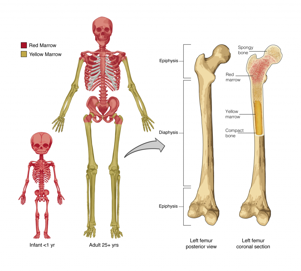
Career Connection – Orthopedist
An orthopedist is a doctor who specializes in diagnosing and treating disorders and injuries related to the musculoskeletal system. Some orthopedic problems can be treated with medications, exercises, braces, and other devices, but others may be best treated with surgery ( Figure 6.1.3 ).

While the origin of the word “orthopedics” (ortho- = “straight”; paed- = “child”), literally means “straightening of the child,” orthopedists can have patients who range from pediatric to geriatric. In recent years, orthopedists have even performed prenatal surgery to correct spina bifida, a congenital defect in which the neural canal in the spine of the fetus fails to close completely during embryologic development.
Orthopedists commonly treat bone and joint injuries but they also treat other bone conditions including curvature of the spine. Lateral curvatures (scoliosis) can be severe enough to slip under the shoulder blade (scapula) forcing it up as a hump. Spinal curvatures can also be excessive dorsoventrally (kyphosis) causing a hunch back and thoracic compression. These curvatures often appear in preteens as the result of poor posture, abnormal growth, or indeterminate causes. Mostly, they are readily treated by orthopedists. As people age, accumulated spinal column injuries and diseases like osteoporosis can also lead to curvatures of the spine, hence the stooping you sometimes see in the elderly.
Some orthopedists sub-specialize in sports medicine, which addresses both simple injuries, such as a sprained ankle, and complex injuries, such as a torn rotator cuff in the shoulder. Treatment can range from exercise to surgery.
Section Review
The major functions of the skeletal system are body support, facilitation of movement, protection of internal organs, storage of minerals and fat, and blood cell formation.
Review Questions
Critical thinking questions.
- Suppose your red bone marrow could not be formed. What functions would your body not be able to perform?
- Suppose your osseous tissue could not store calcium. What functions would your body not be able to perform?
Answers for Critical Thinking Questions
- Without red bone marrow, you would not be able to produce blood cells. The red bone marrow is responsible for forming red and white blood cells as well as platelets. Red blood cells transport oxygen to tissues, and remove carbon dioxide. Without red blood cells, your tissues would not be able to produce ATP using oxygen. White blood cells play a role in the immune system fighting off foreign invaders in our body – without white blood cells you would not be able to recover from infection. Platelets are responsible for clotting your blood when a vessel ruptures. Without platelets you would bleed to death and die.
- The calcium in osseous tissue provides mineral support to bones. Without this calcium, the bones are not rigid and cannot be supportive. The calcium in osseous tissue is also an important storage site, that can release calcium when needed. Other organ systems rely on this calcium for action (specifically, muscle contraction and neural signaling). Without calcium storage, blood calcium levels change dramatically and affect muscle contraction and neural signaling.
This work, Anatomy & Physiology, is adapted from Anatomy & Physiology by OpenStax , licensed under CC BY . This edition, with revised content and artwork, is licensed under CC BY-SA except where otherwise noted.
Images, from Anatomy & Physiology by OpenStax , are licensed under CC BY except where otherwise noted.
Access the original for free at https://openstax.org/books/anatomy-and-physiology/pages/1-introduction .
Anatomy & Physiology Copyright © 2019 by Lindsay M. Biga, Staci Bronson, Sierra Dawson, Amy Harwell, Robin Hopkins, Joel Kaufmann, Mike LeMaster, Philip Matern, Katie Morrison-Graham, Kristen Oja, Devon Quick, Jon Runyeon, OSU OERU, and OpenStax is licensed under a Creative Commons Attribution-ShareAlike 4.0 International License , except where otherwise noted.
Introduction to the Skeletal System
Humans are vertebrates, animals having a vertabral column or backbone . They rely on a sturdy internal frame that is centered on a prominent spine . The human skeletal system consists of bones, cartilage , ligaments and tendons and accounts for about 20 percent of the body weight.
The living bones in our bodies use oxygen and give off waste products in metabolism . They contain active tissues that consume nutrients, require a blood supply and change shape or remodel in response to variations in mechanical stress .
Bones provide a rigid framework, known as the skeleton , that support and protect the soft organs of the body.
The skeleton supports the body against the pull of gravity. The large bones of the lower limbs support the trunk when standing.
The skeleton also protects the soft body parts. The fused bones of the cranium surround the brain to make it less vulnerable to injury. Vertebrae surround and protect the spinal cord and bones of the rib cage help protect the heart and lungs of the thorax.
Bones work together with muscles as simple mechanical lever systems to produce body movement.
Bones contain more calcium than any other organ . The intercellular matrix of bone contains large amounts of calcium salts, the most important being calcium phosphate.
When blood calcium levels decrease below normal, calcium is released from the bones so that there will be an adequate supply for metabolic needs. When blood calcium levels are increased, the excess calcium is stored in the bone matrix. The dynamic process of releasing and storing calcium goes on almost continuously.
Hematopoiesis , the formation of blood cells, mostly takes place in the red marrow of the bones.
In infants, red marrow is found in the bone cavities. With age, it is largely replaced by yellow marrow for fat storage. In adults, red marrow is limited to the spongy bone in the skull , ribs, sternum , clavicles, vertebrae and pelvis . Red marrow functions in the formation of red blood cells, white blood cells and blood platelets.
IMAGES
VIDEO
COMMENTS
In adults, the skeletal system includes 206 bones, many of which are shown in Figure 14.2.2 14.2. 2. Bones are organs made of dense connective tissues, mainly the tough protein collagen. Bones contain blood vessels, nerves, and other tissues. Bones are hard and rigid due to deposits of calcium and other mineral salts within their living tissues.
The vertebral column, or backbone, usually consists of "7 cervical vertebrae, 12 thoracic vertebrae, 5 lumbar vertebrae, 1 sacral bone, and 1 coccyx bone" ( Skeletal System: Bones and Joints 125). It is the central axis of the skeleton that has four major curvatures. Normally, the cervical and the lumbar regions curve anteriorly.
The skeletal system consists of bones, cartilage, and ligaments, which all fall into the category of connective tissue. Bones are classified by their shape. Some are long, some are short, others are flat, and the rest have an irregular shape. There are two types of bone (osseous) tissue. One is cancellous (nicknamed spongy bone due to its ...
A diagram of the human skeleton showing bone and cartilage. Protection of the heart, lungs, and other organs and structures in the chest creates a problem somewhat different from that of the central nervous system. These organs, the function of which involves motion, expansion, and contraction, must have a flexible and elastic protective covering.
The skeletal system provides support and protection to the soft tissues that make up the rest of the body. The skeletal system also provides attachment for muscles to allow movements at the joints, it is a place where to store fat and precious minerals such as calcium and phosphorous. Another function of bones is to house the bone marrow that ...
The skeletal system provides support and protection for the body's internal organs and gives the muscles a point of attachment. Humans have an endoskeleton, where our bones lie underneath our skin and muscles. In other animals, such as insects, there is an exoskeleton on the outside of the body. Overview.
14.2: Introduction to the Skeletal System. The skull and cross-bones symbol has been used for a very long time to represent death, perhaps because after death and decomposition, bones are all that remain. Many people think of bones as being dead, dry, and brittle. These adjectives may correctly describe the bones of a preserved skeleton, but ...
Summary. The skeletal system serves as the frame for the body, providing support and protection. It consists of bones, joints, cartilage, and ligaments. Bones consist of living tissue that undergoes constant remodeling as it matures and ages, whereas joints are the points where two bones meet and can allow for movement.
Unlike a fossil skeleton, however, your skeleton is a structure of living tissue that grows, repairs, and renews itself. The bones within it are dynamic and complex organs that serve a number of important functions, including some necessary to maintain homeostasis. Figure 5.1.1 5.1. 1: Child Looking at Fossils. Bone is a living tissue.
Bone, or osseous tissue, is a hard, dense connective tissue that forms most of the adult skeleton, the support structure of the body. In the areas of the skeleton where bones move (for example, the ribcage and joints), cartilage, a semi-rigid form of connective tissue, provides flexibility and smooth surfaces for movement. The skeletal system ...
1. Introduction The skeletal system consists of bones and their associated connective tissues - including cartilage, tendons, and ligaments. It serves the following functions: • Support: The bones of the legs act as pillars to support the body trunk when standing, and the ribcage supports the thoracic wall. • Protection: Bones surround and protect soft and vital organs. For example, the ...
The human system that consists of bones, cartilage, ligaments, tendons, and joints is the skeletal system. The major functions of this includes protecting organs, allowing movement, and many others. The skeletal system interacts with many other of the human systems such as the circulatory, respiratory, muscular, and immune systems.
The musculoskeletal system. In the musculoskeletal system, the muscular and skeletal systems work together to support and move the body. The bones of the skeletal system serve to protect the body's organs, support the weight of the body, and give the body shape. The muscles of the muscular system attach to these bones, pulling on them to allow ...
The skeletal system gives your body its shape and holds your organs in place. In the simplest terms, your skeletal system is your body's most important support structure. But it's more than just your skeleton and bones. Your skeletal system also includes connective tissue that helps you stay supported and safe while you're moving and still.
The skeletal system, consisting of 206 bones, is one of the most significant systems of the body. Its functions include protecting the organs from damage, supporting the structure of the body, ensuring mobility, and storing minerals. The system maintains homeostasis by producing new blood cells and supplying oxygen and nutrients.
11.2 Summary. The skeletal system is the organ system that provides an internal framework for the human body. In adults, the skeletal system contains 206 bones. Bones are organs made of supportive connective tissues, mainly the tough protein collagen. Bones also contain blood vessels, nerves, and other tissues.
read more (which make up the skeleton), muscles Muscles There are three types of muscles: Skeletal Smooth Cardiac (heart) Two of these kinds—skeletal and smooth—are part of the musculoskeletal system. read more , tendons Tendons and Bursae Tendons are tough bands of connective tissue made up mostly of a rigid protein called collagen ...
The skeletal system function is to form a shield or a solid framework that supports and protects the body's organs and skeletal muscles. Each bone is a complex living organ that is made up of many cells, protein fibers, and minerals. Further more the skeletal system provides attachment points for muscles to allow movements at the joints.
An orthopedist is a doctor who specializes in diagnosing and treating disorders and injuries related to the musculoskeletal system. Some orthopedic problems can be treated with medications, exercises, braces, and other devices, but others may be best treated with surgery (Figure 6.1.3).Figure 6.1.3 - Arm Brace: An orthopedist will sometimes prescribe the use of a brace that reinforces the ...
The skeletal system defines your body shape. It also influences the size of your hands and legs, their height and width. The muscles in the body are attached to the bones, so when they contract together, it causes the bones to move. In long bones, there is a spongy tissue that is responsible for red blood cells.
The skeletal framework of the body is composed of at least 206 bones and the associated tendons, ligaments, and cartilages. The skeletal system has a variety of important functions, including, the support of soft tissues, blood cell production, mineral/electrolyte and lipid storage, and, through its relationships with the muscular system, the support and movement of the body as a whole.
Musculoskeletal system. The musculoskeletal system (locomotor system) is a human body system that provides our body with movement, stability, shape, and support. It is subdivided into two broad systems: Muscular system, which includes all types of muscles in the body. Skeletal muscles, in particular, are the ones that act on the body joints to ...
Introduction to the Skeletal System. Humans are vertebrates, animals having a vertabral column or backbone.They rely on a sturdy internal frame that is centered on a prominent spine.The human skeletal system consists of bones, cartilage, ligaments and tendons and accounts for about 20 percent of the body weight.. The living bones in our bodies use oxygen and give off waste products in metabolism.