Parts of the Brain: Anatomy, Structure & Functions
Olivia Guy-Evans, MSc
Associate Editor for Simply Psychology
BSc (Hons) Psychology, MSc Psychology of Education
Olivia Guy-Evans is a writer and associate editor for Simply Psychology. She has previously worked in healthcare and educational sectors.
Learn about our Editorial Process
Saul Mcleod, PhD
Editor-in-Chief for Simply Psychology
BSc (Hons) Psychology, MRes, PhD, University of Manchester
Saul Mcleod, PhD., is a qualified psychology teacher with over 18 years of experience in further and higher education. He has been published in peer-reviewed journals, including the Journal of Clinical Psychology.
On This Page:
The brain controls all functions of the body, interprets information from the outside world, and defines who we are as individuals and how we experience the world.
The brain receives information through our senses: sight, touch, taste, smell, and hearing. This information is processed in the brain, allowing us to give meaning to the input it receives.
The brain is part of the central nervous system ( CNS ) along with the spinal cord. There is also a peripheral nervous system (PNS) comprised of 31 pairs of spinal nerves that branch from the spinal cord and cranial nerves that branch from the brain.

Brain Parts
The brain is composed of the cerebrum, cerebellum, and brainstem (Fig. 1).

Figure 1. The brain has three main parts: the cerebrum, cerebellum, and brainstem.
The cerebrum is the largest and most recognizable part of the brain. It consists of grey matter (the cerebral cortex ) and white matter at the center. The cerebrum is divided into two hemispheres, the left and right, and contains the lobes of the brain (frontal, temporal, parietal, and occipital lobes).
The cerebrum produces higher functioning roles such as thinking, learning, memory, language, emotion, movement, and perception.
The Cerebellum
The cerebellum is located under the cerebrum and monitors and regulates motor behaviors, especially automatic movements.
This structure is also important for regulating posture and balance and has recently been suggested for being involved in learning and attention.
Although the cerebellum only accounts for roughly 10% of the brain’s total weight, this area is thought to contain more neurons (nerve cells) than the rest of the brain combined.
The brainstem is located at the base of the brain. This area connects the cerebrum and the cerebellum to the spinal cord, acting as a relay station for these areas.
The brainstem regulates automatic functions such as sleep cycles, breathing, body temperature, digestion, coughing, and sneezing.

Right Brain vs. Left Brain
The cerebrum is divided into two halves: the right and left hemispheres (Fig. 2). The left hemisphere controls the right half of the body, and the right hemisphere controls the left half.
The two hemispheres are connected by a thick band of neural fibers known as the corpus callosum, consisting of about 200 million axons.
The corpus callosum allows the two hemispheres to communicate and allows information being processed on one side of the brain to be shared with the other.

Figure 2. The cerebrum is divided into left and right hemispheres. The nerve fibers corpus callosum connects the two sides.
Hemispheric lateralization is the idea that each hemisphere is responsible for different functions. Each of these functions is localized to either the right or left side.
The left hemisphere is associated with language functions, such as formulating grammar and vocabulary and containing different language centers (Broca’s and Wernicke’s area).
The right hemisphere is associated with more visuospatial functions such as visualization, depth perception, and spatial navigation. These left and right functions are the case in most people, especially those who are right-handed.
Lobes of the Brain
Each cerebral hemisphere can be subdivided into four lobes, each associated with different functions.
The four lobes of the brain are the frontal, parietal, temporal, and occipital lobes (Figure 3).

Figure 3. The cerebrum is divided into four lobes: frontal, parietal, occipital, and temporal.
Frontal lobes
The frontal lobes are located at the front of the brain, behind the forehead (Figure 4).
Their main functions are associated with higher cognitive functions, including problem-solving, decision-making, attention, intelligence, and voluntary behaviors.
The frontal lobes contain the motor cortex responsible for planning and coordinating movements.
It also contains the prefrontal cortex, which is responsible for initiating higher-lever cognitive functioning, and Broca’s Area, which is essential for language production.

Figure 4. Frontal lobe structure.
Temporal lobes
The temporal lobes are located on both sides of the brain, near the temples of the head, hence the name temporal lobes (Figure 5).
The main functions of these lobes include understanding, language, memory acquisition, face recognition, object recognition, perception, and auditory information processing.
There is a temporal lobe in both the left and right hemispheres. The left temporal lobe, which is usually the most dominant in people, is associated with language, learning, memorizing, forming words, and remembering verbal information.
The left lobe also contains a vital language center known as Wernicke’s area, which is essential for language development. The right temporal lobe is usually associated with learning and memorizing non-verbal information and determining facial expressions.

Figure 5. Temporal lobe structure.
Parietal lobes
The parietal lobe is located at the top of the brain, between the frontal and occipital lobes, and above the temporal lobes (Figure 6).
The parietal lobe is essential for integrating information from the body’s senses to allow us to build a coherent picture of the world around us.
These lobes allow us to perceive our bodies through somatosensory information (e.g., through touch, pressure, and temperature). It can also help with visuospatial processing, reading, and number representations (mathematics).
The parietal lobes also contain the somatosensory cortex, which receives and processes sensory information, integrating this into a representational map of the body.
This means it can pinpoint the exact area of the body where a sensation is felt, as well as perceive the weight of objects, shape, and texture.

Figure 6. Parietal lobe structure.
Occipital lobes
The occipital lobes are located at the back of the brain behind the temporal and parietal lobes and below the occipital bone of the skull (Figure 7).
The occipital lobes receive sensory information from the eyes’ retinas, which is then encoded into different visual data. Some of the functions of the occipital lobes include being able to assess the size, depth, and distance, determine color information, object and facial recognition, and mapping the visual world.
The occipital lobes also contain the primary visual cortex, which receives sensory information from the retinas, transmitting this information relating to location, spatial data, motion, and the colors of objects in the field of vision.

Figure 7. Occipital lobe structure.
Cerebral Cortex
The surface of the cerebrum is called the cerebral cortex and has a wrinkled appearance, consisting of bulges, also known as gyri, and deep furrows, known as sulci (Figure 8).
A gyrus (plural: gyri) is the name given to the bumps and ridges on the cerebral cortex (the outermost layer of the brain). A sulcus (plural: sulci) is another name for a groove in the cerebral cortex.

Figure 8. The cortex contains neurons (grey matter) interconnected to other brain areas by axons (white matter). The cortex has a folded appearance. A fold is called a gyrus, and the valley between is a sulcus.
The cerebral cortex is primarily constructed of grey matter (neural tissue made up of neurons), with between 14 and 16 billion neurons found here.
The many folds and wrinkles of the cerebral cortex allow a wider surface area for an increased number of neurons to live there, permitting large amounts of information to be processed.
Deep Structures
The amygdala is a structure deep in the brain that is involved in the processing of emotions and fear learning. The amygdala is a part of the limbic system, a neural network that mediates emotion and memory (Figure 9).
This structure also ties emotional meaning to memories, processes rewards, and helps us make decisions. This structure has also been linked with the fight-or-flight response.

Figure 9. The amygdala in the limbic system plays a key role in how animals assess and respond to environmental threats and challenges by evaluating the emotional importance of sensory information and prompting an appropriate response.
Thalamus and Hypothalamus
The thalamus relays information between the cerebral cortex, brain stem, and other cortical structures (Figure 10).
Because of its interactive role in relaying sensory and motor information, the thalamus contributes to many processes, including attention, perception, timing, and movement. The hypothalamus modulates a range of behavioral and physiological functions.
It controls autonomic functions such as hunger, thirst, body temperature, and sexual activity. To do this, the hypothalamus integrates information from different brain parts and responds to various stimuli such as light, odor, and stress.

Figure 10. The thalamus is often described as the brain’s relay station as a great deal of information that reaches the cerebral cortex first stops in the thalamus before being sent to its destination.
Hippocampus
The hippocampus is a curved-shaped structure in the limbic system associated with learning and memory (Figure 11).
This structure is most strongly associated with the formation of memories, is an early storage system for new long-term memories, and plays a role in the transition of these long-term memories to more permanent memories.

Figure 11. Hippocampus location in the brain
Basal Ganglia
The basal ganglia are a group of structures that regulate the coordination of fine motor movements, balance, and posture alongside the cerebellum.
These structures are connected to other motor areas and link the thalamus with the motor cortex. The basal ganglia are also involved in cognitive and emotional behaviors, as well as playing a role in reward and addiction.

Figure 12. The Basal Ganglia Illustration
Ventricles and Cerebrospinal Fluid
Within the brain, there are fluid-filled interconnected cavities called ventricles , which are extensions of the spinal cord. These are filled with a substance called cerebrospinal fluid, which is a clear and colorless liquid.
The ventricles produce cerebrospinal fluid and transport and remove this fluid. The ventricles do not have a unique function, but they provide cushioning to the brain and are useful for determining the locations of other brain structures.
Cerebrospinal fluid circulates the brain and spinal cord and functions to cushion the brain within the skull. If damage occurs to the skull, the cerebrospinal fluid will act as a shock absorber to help protect the brain from injury.

As well as providing cushioning, the cerebrospinal fluid circulates nutrients and chemicals filtered from the blood and removes waste products from the brain. Cerebrospinal fluid is constantly absorbed and replenished by the ventricles.
If there were a disruption or blockage, this can cause a build-up of cerebrospinal fluid and can cause enlarged ventricles.
Neurons are the nerve cells of the central nervous system that transmit information through electrochemical signals throughout the body. Neurons contain a soma, a cell body from which the axon extends.
Axons are nerve fibers that are the longest part of the neuron, which conduct electrical impulses away from the soma.

There are dendrites at the end of the neuron, which are branch-like structures that send and receive information from other neurons.
A myelin sheath, a fatty insulating layer, forms around the axon, allowing nerve impulses to travel down the axon quickly.
There are different types of neurons. Sensory neurons transmit sensory information, motor neurons transmit motor information, and relay neurons allow sensory and motor neurons to communicate.
The communication between neurons is called synapses. Neurons communicate with each other via synaptic clefts, which are gaps between the endings of neurons.

During synaptic transmission, chemicals, such as neurotransmitters, are released from the endings of the previous neuron (also known as the presynaptic neuron).
These chemicals enter the synaptic cleft to then be transported to receptors on the next neuron (also known as the postsynaptic neuron).
Once transported to the next neuron, the chemical messengers continue traveling down neurons to influence many functions, such as behavior and movement.
Glial Cells
Glial cells are non-neuronal cells in the central nervous system which work to provide the neurons with nourishment, support, and protection.
These are star-shaped cells that function to maintain the environment for neuronal signaling by controlling the levels of neurotransmitters surrounding the synapses.
They also work to clean up what is left behind after synaptic transmission, either recycling any leftover neurotransmitters or cleaning up when a neuron dies.
Oligodendrocytes
These types of glial have the appearance of balls with spikes all around them. They function by wrapping around the axons of neurons to form a protective layer called the myelin sheath.
This is a substance that is rich in fat and provides insulation to the neurons to aid neuronal signaling.
Microglial cells have oval bodies and many branches projecting out of them. The primary function of these cells is to respond to injuries or diseases in the central nervous system.
They respond by clearing away any dead cells or removing any harmful toxins or pathogens that may be present, so they are, therefore, important to the brain’s health.
Ependymal cells
These cells are column-shaped and usually line up together to form a membrane called the ependyma. The ependyma is a thin membrane lining the spinal cord and ventricles of the brain .
In the ventricles, these cells have small hairlike structures called cilia, which help encourage the flow of cerebrospinal fluid.
Cranial Nerves
There are 12 types of cranial nerves which are linked directly to the brain without having to pass through the spinal cord. These allow sensory information to pass from the organs of the face to the brain:

Mnemonic for Order of Cranial Nerves:
S ome S ay M arry M oney B ut M y B rother S ays B ig B rains M atter M ore
- Cranial I: Sensory
- Cranial II: Sensory
- Cranial III: Motor
- Cranial IV: Motor
- Cranial V: Both (sensory & motor)
- Cranial VI: Motor
- Cranial VII: Both (sensory & motor)
- Cranial VIII: Sensory
- Cranial IX: Both (sensory & motor)
- Cranial X: Both (sensory & motor)
- Cranial XI: Motor
- Cranial XII: Motor
Purves, D., Augustine, G., Fitzpatrick, D., Katz, L., LaMantia, A., McNamara, J., & Williams, S. (2001). Neuroscience 2nd edition . sunderland (ma) sinauer associates. Types of Eye Movements and Their Functions.
Mayfield Brain and Spine (n.d.). Anatomy of the Brain. Retrieved July 28, 2021, from: https://mayfieldclinic.com/pe-anatbrain.htm
Robertson, S. (2018, August 23). What is Grey Matter? News Medical Life Sciences. https://www.news-medical.net/health/What-is-Grey-Matter.aspx
Guy-Evans, O. (2021, April 13). Temporal lobe: definition, functions, and location. Simply Psychology. www.www.www.www.www.www.simplypsychology.org/temporal-lobe.html
Guy-Evans, O. (2021, April 15). Parietal lobe: definition, functions, and location. Simply Psychology. www.www.www.www.www.www.simplypsychology.org/parietal-lobe.html
Guy-Evans, O. (2021, April 19). Occipital lobe: definition, functions, and location. Simply Psychology. www.www.www.www.www.www.simplypsychology.org/occipital-lobe.html
Guy-Evans, O. (2021, May 08). Frontal lobe function, location in brain, damage, more. Simply Psychology. www.www.www.www.www.www.simplypsychology.org/frontal-lobe.html
Guy-Evans, O. (2021, June 09). Gyri and sulci of the brain. Simply Psychology. www.www.www.www.www.www.simplypsychology.org/gyri-and-sulci-of-the-brain.html

Masks Strongly Recommended but Not Required in Maryland, Starting Immediately
Due to the downward trend in respiratory viruses in Maryland, masking is no longer required but remains strongly recommended in Johns Hopkins Medicine clinical locations in Maryland. Read more .
- Vaccines
- Masking Guidelines
- Visitor Guidelines

Brain Anatomy and How the Brain Works
What is the brain.
The brain is a complex organ that controls thought, memory, emotion, touch, motor skills, vision, breathing, temperature, hunger and every process that regulates our body. Together, the brain and spinal cord that extends from it make up the central nervous system, or CNS.
What is the brain made of?
Weighing about 3 pounds in the average adult, the brain is about 60% fat. The remaining 40% is a combination of water, protein, carbohydrates and salts. The brain itself is a not a muscle. It contains blood vessels and nerves, including neurons and glial cells.
What is the gray matter and white matter?
Gray and white matter are two different regions of the central nervous system. In the brain, gray matter refers to the darker, outer portion, while white matter describes the lighter, inner section underneath. In the spinal cord, this order is reversed: The white matter is on the outside, and the gray matter sits within.

Gray matter is primarily composed of neuron somas (the round central cell bodies), and white matter is mostly made of axons (the long stems that connects neurons together) wrapped in myelin (a protective coating). The different composition of neuron parts is why the two appear as separate shades on certain scans.

Each region serves a different role. Gray matter is primarily responsible for processing and interpreting information, while white matter transmits that information to other parts of the nervous system.
How does the brain work?
The brain sends and receives chemical and electrical signals throughout the body. Different signals control different processes, and your brain interprets each. Some make you feel tired, for example, while others make you feel pain.
Some messages are kept within the brain, while others are relayed through the spine and across the body’s vast network of nerves to distant extremities. To do this, the central nervous system relies on billions of neurons (nerve cells).
Main Parts of the Brain and Their Functions
At a high level, the brain can be divided into the cerebrum, brainstem and cerebellum.

The cerebrum (front of brain) comprises gray matter (the cerebral cortex) and white matter at its center. The largest part of the brain, the cerebrum initiates and coordinates movement and regulates temperature. Other areas of the cerebrum enable speech, judgment, thinking and reasoning, problem-solving, emotions and learning. Other functions relate to vision, hearing, touch and other senses.
Cerebral Cortex
Cortex is Latin for “bark,” and describes the outer gray matter covering of the cerebrum. The cortex has a large surface area due to its folds, and comprises about half of the brain’s weight.
The cerebral cortex is divided into two halves, or hemispheres. It is covered with ridges (gyri) and folds (sulci). The two halves join at a large, deep sulcus (the interhemispheric fissure, AKA the medial longitudinal fissure) that runs from the front of the head to the back. The right hemisphere controls the left side of the body, and the left half controls the right side of the body. The two halves communicate with one another through a large, C-shaped structure of white matter and nerve pathways called the corpus callosum. The corpus callosum is in the center of the cerebrum.
The brainstem (middle of brain) connects the cerebrum with the spinal cord. The brainstem includes the midbrain, the pons and the medulla.
- Midbrain. The midbrain (or mesencephalon) is a very complex structure with a range of different neuron clusters (nuclei and colliculi), neural pathways and other structures. These features facilitate various functions, from hearing and movement to calculating responses and environmental changes. The midbrain also contains the substantia nigra, an area affected by Parkinson’s disease that is rich in dopamine neurons and part of the basal ganglia, which enables movement and coordination.
- Pons. The pons is the origin for four of the 12 cranial nerves, which enable a range of activities such as tear production, chewing, blinking, focusing vision, balance, hearing and facial expression. Named for the Latin word for “bridge,” the pons is the connection between the midbrain and the medulla.
- Medulla. At the bottom of the brainstem, the medulla is where the brain meets the spinal cord. The medulla is essential to survival. Functions of the medulla regulate many bodily activities, including heart rhythm, breathing, blood flow, and oxygen and carbon dioxide levels. The medulla produces reflexive activities such as sneezing, vomiting, coughing and swallowing.
The spinal cord extends from the bottom of the medulla and through a large opening in the bottom of the skull. Supported by the vertebrae, the spinal cord carries messages to and from the brain and the rest of the body.
The cerebellum (“little brain”) is a fist-sized portion of the brain located at the back of the head, below the temporal and occipital lobes and above the brainstem. Like the cerebral cortex, it has two hemispheres. The outer portion contains neurons, and the inner area communicates with the cerebral cortex. Its function is to coordinate voluntary muscle movements and to maintain posture, balance and equilibrium. New studies are exploring the cerebellum’s roles in thought, emotions and social behavior, as well as its possible involvement in addiction, autism and schizophrenia.
Brain Coverings: Meninges
Three layers of protective covering called meninges surround the brain and the spinal cord.
- The outermost layer, the dura mater , is thick and tough. It includes two layers: The periosteal layer of the dura mater lines the inner dome of the skull (cranium) and the meningeal layer is below that. Spaces between the layers allow for the passage of veins and arteries that supply blood flow to the brain.
- The arachnoid mater is a thin, weblike layer of connective tissue that does not contain nerves or blood vessels. Below the arachnoid mater is the cerebrospinal fluid, or CSF. This fluid cushions the entire central nervous system (brain and spinal cord) and continually circulates around these structures to remove impurities.
- The pia mater is a thin membrane that hugs the surface of the brain and follows its contours. The pia mater is rich with veins and arteries.

Lobes of the Brain and What They Control
Each brain hemisphere (parts of the cerebrum) has four sections, called lobes: frontal, parietal, temporal and occipital. Each lobe controls specific functions.

- Frontal lobe. The largest lobe of the brain, located in the front of the head, the frontal lobe is involved in personality characteristics, decision-making and movement. Recognition of smell usually involves parts of the frontal lobe. The frontal lobe contains Broca’s area, which is associated with speech ability.
- Parietal lobe. The middle part of the brain, the parietal lobe helps a person identify objects and understand spatial relationships (where one’s body is compared with objects around the person). The parietal lobe is also involved in interpreting pain and touch in the body. The parietal lobe houses Wernicke’s area, which helps the brain understand spoken language.
- Occipital lobe. The occipital lobe is the back part of the brain that is involved with vision.
- Temporal lobe. The sides of the brain, temporal lobes are involved in short-term memory, speech, musical rhythm and some degree of smell recognition.
Deeper Structures Within the Brain
Pituitary gland.
Sometimes called the “master gland,” the pituitary gland is a pea-sized structure found deep in the brain behind the bridge of the nose. The pituitary gland governs the function of other glands in the body, regulating the flow of hormones from the thyroid, adrenals, ovaries and testicles. It receives chemical signals from the hypothalamus through its stalk and blood supply.
Hypothalamus
The hypothalamus is located above the pituitary gland and sends it chemical messages that control its function. It regulates body temperature, synchronizes sleep patterns, controls hunger and thirst and also plays a role in some aspects of memory and emotion.
Small, almond-shaped structures, an amygdala is located under each half (hemisphere) of the brain. Included in the limbic system, the amygdalae regulate emotion and memory and are associated with the brain’s reward system, stress, and the “fight or flight” response when someone perceives a threat.
Hippocampus
A curved seahorse-shaped organ on the underside of each temporal lobe, the hippocampus is part of a larger structure called the hippocampal formation. It supports memory, learning, navigation and perception of space. It receives information from the cerebral cortex and may play a role in Alzheimer’s disease.
Pineal Gland
The pineal gland is located deep in the brain and attached by a stalk to the top of the third ventricle. The pineal gland responds to light and dark and secretes melatonin, which regulates circadian rhythms and the sleep-wake cycle.
Ventricles and Cerebrospinal Fluid
Deep in the brain are four open areas with passageways between them. They also open into the central spinal canal and the area beneath arachnoid layer of the meninges.
The ventricles manufacture cerebrospinal fluid , or CSF, a watery fluid that circulates in and around the ventricles and the spinal cord, and between the meninges. CSF surrounds and cushions the spinal cord and brain, washes out waste and impurities, and delivers nutrients.

Blood Supply to the Brain
Two sets of blood vessels supply blood and oxygen to the brain: the vertebral arteries and the carotid arteries.
The external carotid arteries extend up the sides of your neck, and are where you can feel your pulse when you touch the area with your fingertips. The internal carotid arteries branch into the skull and circulate blood to the front part of the brain.
The vertebral arteries follow the spinal column into the skull, where they join together at the brainstem and form the basilar artery , which supplies blood to the rear portions of the brain.
The circle of Willis , a loop of blood vessels near the bottom of the brain that connects major arteries, circulates blood from the front of the brain to the back and helps the arterial systems communicate with one another.

Cranial Nerves
Inside the cranium (the dome of the skull), there are 12 nerves, called cranial nerves:
- Cranial nerve 1: The first is the olfactory nerve, which allows for your sense of smell.
- Cranial nerve 2: The optic nerve governs eyesight.
- Cranial nerve 3: The oculomotor nerve controls pupil response and other motions of the eye, and branches out from the area in the brainstem where the midbrain meets the pons.
- Cranial nerve 4: The trochlear nerve controls muscles in the eye. It emerges from the back of the midbrain part of the brainstem.
- Cranial nerve 5: The trigeminal nerve is the largest and most complex of the cranial nerves, with both sensory and motor function. It originates from the pons and conveys sensation from the scalp, teeth, jaw, sinuses, parts of the mouth and face to the brain, allows the function of chewing muscles, and much more.
- Cranial nerve 6: The abducens nerve innervates some of the muscles in the eye.
- Cranial nerve 7: The facial nerve supports face movement, taste, glandular and other functions.
- Cranial nerve 8: The vestibulocochlear nerve facilitates balance and hearing.
- Cranial nerve 9: The glossopharyngeal nerve allows taste, ear and throat movement, and has many more functions.
- Cranial nerve 10: The vagus nerve allows sensation around the ear and the digestive system and controls motor activity in the heart, throat and digestive system.
- Cranial nerve 11: The accessory nerve innervates specific muscles in the head, neck and shoulder.
- Cranial nerve 12: The hypoglossal nerve supplies motor activity to the tongue.
The first two nerves originate in the cerebrum, and the remaining 10 cranial nerves emerge from the brainstem, which has three parts: the midbrain, the pons and the medulla.
Find a Treatment Center
- Neurology and Neurosurgery
Find Additional Treatment Centers at:
- Howard County Medical Center
- Sibley Memorial Hospital
- Suburban Hospital
Request an Appointment

Facioscapulohumeral Muscular Dystrophy in Children

Moyamoya Disease
Motor Stereotypies
Related Topics

- Science Notes Posts
- Contact Science Notes
- Todd Helmenstine Biography
- Anne Helmenstine Biography
- Free Printable Periodic Tables (PDF and PNG)
- Periodic Table Wallpapers
- Interactive Periodic Table
- Periodic Table Posters
- How to Grow Crystals
- Chemistry Projects
- Fire and Flames Projects
- Holiday Science
- Chemistry Problems With Answers
- Physics Problems
- Unit Conversion Example Problems
- Chemistry Worksheets
- Biology Worksheets
- Periodic Table Worksheets
- Physical Science Worksheets
- Science Lab Worksheets
- My Amazon Books
Parts of the Brain and Their Functions

The human brain is the epicenter of our nervous system and plays a pivotal role in virtually every aspect of our lives. It’s a complex, highly organized organ responsible for thoughts, feelings, actions, and interactions with the world around us. Here is a look at the intricate anatomy of the brain, its functions, and the consequences of damage to different areas.
Introduction to the Brain and Its Functions
The brain is an organ of soft nervous tissue that is protected within the skull of vertebrates. It functions as the coordinating center of sensation and intellectual and nervous activity. The brain consists of billions of neurons (nerve cells) that communicate through intricate networks. The primary functions of the brain include processing sensory information, regulating bodily functions, forming thoughts and emotions, and storing memories.
Main Parts of the Brain – Anatomy
The three main parts of the brain are the cerebrum, cerebellum, and brainstem.
1. Cerebrum
- Location: The cerebellum occupies the upper part of the cranial cavity and is the largest part of the human brain.
- Functions: It’s responsible for higher brain functions, including thought, action, emotion, and interpretation of sensory data.
- Effects of Damage: Depending on the area affected, damage leads to memory loss, impaired cognitive skills, changes in personality, and loss of motor control.
2. Cerebellum
- Location: The cerebellum is at the back of the brain, below the cerebrum.
- Functions: It coordinates voluntary movements such as posture, balance, coordination, and speech.
- Effects of Damage: Damage causes problems with balance, movement, and muscle coordination (ataxia).
3. Brainstem
- Location: The brainstem is lower extension of the brain, connecting to the spinal cord. It includes the midbrain, pons, and medulla oblongata.
- Functions: This part of the brain controls many basic life-sustaining functions, including heart rate, breathing, sleeping, and eating.
- Effects of Damage: Damage results in life-threatening conditions like breathing difficulties, heart problems, and loss of consciousness.
Lobes of the Brain
The four lobes of the brain are regions of the cerebrum:
- Location: This is the anterior or front part of the brain.
- Functions: Decision making, problem solving, control of purposeful behaviors, consciousness, and emotions.
- Location: Sits behind the frontal lobe.
- Functions: Processes sensory information it receives from the outside world, mainly relating to spatial sense and navigation (proprioception).
- Location: Below the lateral fissure, on both cerebral hemispheres.
- Functions: Mainly revolves around auditory perception and is also important for the processing of both speech and vision (reading).
- Location: At the back of the brain.
- Functions: Main center for visual processing.
Left vs. Right Brain Hemispheres
The cerebrum has two halves, called hemispheres. Each half controls functions on the opposite side of the body. So, the left hemisphere controls muscles on the right side of the body, and vice versa. But, the functions of the two hemispheres are not entirely identical:
- Left Hemisphere: It’s dominant in language and speech and plays roles in logical thinking, analysis, and accuracy. .
- Right Hemisphere: This hemisphere is more visual and intuitive and functions in creative and imaginative tasks.
The corpus callosum is a band of nerves that connect the two hemispheres and allow communication between them.
Detailed List of Parts of the Brain
While knowing the three key parts of the brain is a good start, the anatomy is quite a bit more complex. In addition to nervous tissues, the brain also contains key glands:
- Cerebrum: The cerebrum is the largest part of the brain. Divided into lobes, it coordinates thought, movement, memory, senses, speech, and temperature.
- Corpus Callosum : A broad band of nerve fibers joining the two hemispheres of the brain, facilitating interhemispheric communication.
- Cerebellum : Coordinates movement and balance and aids in eye movement.
- Pons : Controls voluntary actions, including swallowing, bladder function, facial expression, posture, and sleep.
- Medulla oblongata : Regulates involuntary actions, including breathing, heart rhythm, as well as oxygen and carbon dioxide levels.
- Limbic System : Includes the amygdala, hippocampus, and parts of the thalamus and hypothalamus.
- Amygdala: Plays a key role in emotional responses, hormonal secretions, and memory formation.
- Hippocampus: Plays a vital role in memory formation and spatial navigation.
- Thalamus : Acts as the brain’s relay station, channeling sensory and motor signals to the cerebral cortex, and regulating consciousness, sleep, and alertness.
- Basal Ganglia : A group of structures involved in processing information related to movement, emotions, and reward. Key structures include the striatum, globus pallidus, substantia nigra, and subthalamic nucleus.
- Ventral Tegmental Area (VTA) : Plays a role in the reward circuit of the brain, releasing dopamine in response to stimuli indicating a reward.
- Optic tectum : Also known as the superior colliculus, it directs eye movements.
- Substantia Nigra : Involved in motor control and contains a large concentration of dopamine-producing neurons.
- Cingulate Gyrus : Plays a role in processing emotions and behavior regulation. It also helps regulate autonomic motor function.
- Olfactory Bulb : Involved in the sense of smell and the integration of olfactory information.
- Mammillary Bodies : Plays a role in recollective memory.
- Function: Regulates emotions, memory, and arousal.
Glands in the Brain
The hypothalamus, pineal gland, and pituitary gland are the three endocrine glands within the brain:
- Hypothalamus : The hypothalamus links the nervous and endocrine systems. It contains many small nuclei. In addition to participating in eating and drinking, sleeping and waking, it regulates the endocrine system via the pituitary gland. It maintains the body’s homeostasis, regulating hunger, thirst, response to pain, levels of pleasure, sexual satisfaction, anger, and aggressive behavior.
- Pituitary Gland : Known as the “master gland,” it controls various other hormone glands in the body, such as the thyroid and adrenals, as well as regulating growth, metabolism, and reproductive processes.
- Pineal Gland : The pineal gland produces and regulates some hormones, including melatonin, which is crucial in regulating sleep patterns and circadian rhythms.
Gray Matter vs. White Matter
The brain and spinal cord consist of gray matter (substantia grisea) and white matter (substantia alba).
- White Matter: Consists mainly of axons and myelin sheaths that send signals between different brain regions and between the brain and spinal cord.
- Gray Matter: Consists of neuronal cell bodies, dendrites, and axon terminals. Gray matter processes information and directs stimuli for muscle control, sensory perception, decision making, and self-control.
Frequently Asked Questions (FAQs) About the Human Brain
- The human brain contains approximately 86 billion neurons. Additionally, it has a similar or slightly higher number of non-neuronal cells (glial cells), making the total number of cells in the brain close to 170 billion.
- There are about 86 billion neurons in the human brain. These neurons are connected by trillions of synapses, forming a complex networks.
- The average adult human brain weighs about 1.3 to 1.4 kilograms (about 3 pounds). This weight represents about 2% of the total body weight.
- The brain is about 73% water.
- The myth that humans only use 10% of their brain is false. Virtually every part gets use, and most of the brain is active all the time, even during sleep.
- The average size of the adult human brain is about 15 centimeters (6 inches) in length, 14 centimeters (5.5 inches) in width, and 9 centimeters (3.5 inches) in height.
- Brain signal speeds vary depending on the type of neuron and the nature of the signal. They travel anywhere from 1 meter per second to over 100 meters per second in the fastest neurons.
- With age, the brain’s volume and/or weight decrease, synaptic connections reduce, and there can be a decline in cognitive functions. However, the brain to continues adapting and forming new connections throughout life.
- The brain has a limited ability to repair itself. Neuroplasticity aids recovery by allowing other parts of the brain to take over functions of the damaged areas.
- The brain consumes about 20% of the body’s total energy , despite only making up about 2% of the body’s total weight . It requires a constant supply of glucose and oxygen.
- Sleep is crucial for brain health. It aids in memory consolidation, learning, brain detoxification, and the regulation of mood and cognitive functions.
- Douglas Fields, R. (2008). “White Matter Matters”. Scientific American . 298 (3): 54–61. doi: 10.1038/scientificamerican0308-54
- Kandel, Eric R.; Schwartz, James Harris; Jessell, Thomas M. (2000). Principles of Neural Science (4th ed.). New York: McGraw-Hill. ISBN 978-0-8385-7701-1.
- Kolb, B.; Whishaw, I.Q. (2003). Fundamentals of Human Neuropsychology (5th ed.). New York: Worth Publishing. ISBN 978-0-7167-5300-1.
- Rajmohan, V.; Mohandas, E. (2007). “The limbic system”. Indian Journal of Psychiatry . 49 (2): 132–139. doi: 10.4103/0019-5545.33264
- Shepherd, G.M. (1994). Neurobiology . Oxford University Press. ISBN 978-0-19-508843-4.
Related Posts
Choose Your Test
Sat / act prep online guides and tips, the 3 major parts of the brain and what they do.
General Education

Mission control. Command center. Control tower. No, I'm not talking about space or your laptop hard drive, or even airport flight control. I'm talking about the human brain—the most complex and essential organ our bodies have. What is the brain structure? What part of the brain controls emotions?
Whether you're studying it in class, preparing for an AP exam, or just curious about brain structure, in this article, you'll learn about the main parts of brain anatomy and their functions and as well as get a general overview of the brain's supporting cast.
What Is the Brain and Why Does It Matter?
The brain is a three-pound organ that serves as headquarters for our bodies. Without it, we wouldn't be able to process information, move our limbs, or even breathe . Together with the spinal cord, brain structure and function helps control the central nervous system—the main part of two that make up the human nervous system. (The other part, the peripheral nervous system, is made up of nerves and neurons that connect the central nervous system to the body's limbs and organs.) The human nervous system is responsible for helping us think, breathe, move, react and feel.

Like any good command center, there is a structure to the brain and its operations that help it carry out its basic functions.
What Are the Main Parts of the Brain?
There are three main parts of the brain: the cerebrum, cerebellum and the brain stem.

Image source: Denise Wawrzyniak , used under CC BY-NC 4.0
What Is the Cerebrum?
Was I A Bee /Wikimedia Commons
The cerebrum is the largest part of the brain. Located in the front and middle part of the brain, it accounts for 85% of the brain's weight. Of the three main parts of the brain, the cerebrum is considered the most recent to develop in human evolution. The cerebrum is responsible for all voluntary actions (e.g.: motor skills), communication, emotions, creativity, intelligence and personality.
What Are the Main Parts of the Cerebrum?
The cerebrum's structure is made up of:

What Are the Layers of the Cerebrum?
The cerebrum has two layers: one inner and one outer . The outer layer is known as the cerebral cortex (represented in red in the spinning image above). Most times, whenever you see photos of the brain, you are looking at the cerebral cortex. This area houses the brain's "gray matter," and is considered the "seat" of human consciousness. Higher brain functions such as thinking, reasoning, planning, emotion, memory, the processing of sensory information and speech all happen in the cerebral cortex. In other words, the cerebral cortex is what sets humans apart from other species.

The cerebral cortex is referred to as "gray matter," due to its color and is responsible for several vital functions, such as those listed above.
What Is the Corpus Callosum?
The cerebrum's inner core houses the brain's "white matter." The major part of the inner core is known as the corpus callosum. The corpus callosum is a thick tract of fibrous nerves that serve as a kind of switchboard enabling the brain's hemispheres to communicate with one another. Whereas the cerebral cortex is the cerebrum's outer layer made up of gray matter, and is responsible for thinking, motor function and information processing; the corpus callosum is the cerebrum's inner core, made up of white matter, with four parts of nerve tracts connecting to different parts of the hemispheres.
Home of the white matter: corpus callosum./ Life Sciences Database /Wikimedia Commons
The corpus callosum's nerve fibers (or axons) are coated with myelin. This fatty substance helps increase the transmission of information between the next part of the cerebrum: the two hemispheres.
The Cerebrum's Left and Right Hemispheres
In addition to two layers, the cerebrum also has two halves, or hemispheres: the left hemisphere and the right hemisphere. Although each hemisphere is known for managing different functions, it is important to note that both handle most processes of the brain structure.
The existence of the hemispheres is vital to our body's functions. The relationship between our brain and body is contralateral. This means, generally speaking, that the left side of the brain (left hemisphere) controls the right side of the body, and the right side of the brain (right hemisphere) controls the left side of the body. Because the hemispheres carry out different tasks, they need to "talk" to one another in real time to coordinate our movements, thoughts, etc.
The left hemisphere is responsible for controlling the right side of the body. It handles language, reasoning, logic and speech. If the left hemisphere were a set of classes in school, it would be your math, science, and English classes.
The right hemisphere is responsible for controlling the left side of the body. It handles spatially-related tasks and visual understanding. In terms of classes, the right hemisphere would be your arts, music, and creative writing classes.

The deep groove separating the hemispheres is called the longitudinal fissure (or, cerebral fissure).
The longitudinal fissure is prevented from completely splitting the cerebrum in half by the corpus callosum. Thanks to the corpus callosum (our brain's speedy switchboard), the left side of your brain can chat instantaneously with the right side of your brain.
The red line down the center of the cerebrum is the longitudinal fissure. Life Sciences Database /Wikimedia Commons
All this hemisphere talk brings us to the final part of the cerebrum: the four lobes.
What Are the 4 Lobes of the Brain?
Database Center for Life Sciences /Wikimedia Commons
The cerebrum's left and right hemispheres are each divided into four lobes: the frontal, parietal, occipital and temporal lobes. The lobes generally handle different functions, but much like the hemispheres, the lobes don't function alone. The lobes are separated from each other by depressions in the cortex known as sulcus (or sulci) and are protected by the skull with bones named after their corresponding lobes.

Cancer Research UK /Wikimedia Commons
The frontal lobe is located in the front of the brain, running from your forehead to your ears. It is responsible for problem-solving and planning, thought, behavior, speech, memory and movement. The frontal lobe is separated from the parietal lobe by the central sulcus and is protected by a singular frontal skull bone.
The parietal lobe picks up where the frontal lobe ends and goes until the mid-back part of the brain (about where a ponytail would be). It is responsible for processing information from the senses (touch, sight, hearing, smelling and sight), as well as language interpretation and spatial perception. It is separated from the other lobes on all four sides: from the frontal lobe by central sulcus; from the opposite hemisphere by the longitudinal fissure; from the occipital lobe by parieto-occipital sulcus; and from the temporal lobe below by a depression known as the lateral sulcus, or lateral fissure. Because each hemisphere has a parietal lobe, there are two parietal skull bones—one on the external side of each hemisphere.
The occipital lobe is located in the back of the brain. It is considered the brain's "visual processing center" because it's where the bulk of information our eyes take in gets analyzed and sorted. It is separated from the parietal lobe by the parieto-occipital sulcus; from the temporal lobe by the lateral occipital sulcus; and from the cerebellum (the second part of the brain, coming up soon) by what is called the cerebellar tentorium (or tentorium cerebelli). It is protected by the skull's singular occipital bone.
The temporal lobe is located in behind and below the frontal lobe (and beneath the parietal lobe), under the lateral fissure. It is responsible for our memory, emotions, language and speech, and auditory and visual processing. It is separated from the parietal and frontal lobe by the lateral sulcus (lateral fissure); from the occipital lobe by the lateral occipital sulcus and occipitotemporal sulcus; and is adjacent to the corpus callosum. Similar to its parietal neighbor, the temporal lobe is protected by two bones—one temporal bone on the external side of each hemisphere.

The brain's lobes serve as a map for understanding where brain functions happen. Image source: Denise Wawrzyniak , used under CC BY-NC 4.0
What Is the Cerebellum?
The cerebellum stands for "little brain" in Latin. It looks like a separate mini-brain behind and underneath the cerebrum (beneath the temporal and occipital lobes) and above the brain stem. The cerebellum (along with the brain stem) is considered evolutionarily to be the oldest part of the brain.
Where the cerebrum makes up 85% of the brain's mass, the cerebellum takes up only 10%. However, the cerebellum accounts for more than half of the brain's neurons. The cerebellum is responsible for voluntary movements, coordination, balance, posture, muscle tone, and cognitive functions.
What Are the Main Parts of the Cerebellum?
The cerebellum's structure is made up of:

The Cerebellum's Inner and Outer Layers
Like the cerebrum, the cerebellum has two layers: one inner and one outer. The outer layer is called the cerebellar cortex. Like the cerebral cortex, it is full of gray matter. Functions such as movement, motor learning, balance and posture happen here.
Underneath the cortex lies the cerebellum's white matter. Called "arbor vitae" ("tree of life") for its appearance, the cerebellum's white matter contains cerebellar nuclei. These neurons are vital because they relay information between the cerebral cortex and the peripheral nervous system to assist in learning and cognitive functions, motor control, balance and coordination. (In a very loose parallel, both the cerebellar nuclei of the cerebellum and the corpus callosum of the cerebrum are responsible for internal communications: The corpus callosum relays messages between there cerebrum's hemispheres, and the cerebellar nuclei relays messages between the body and cerebrum.)
The Cerebellum's Left and Right Hemispheres
The cerebellum also has two hemispheres: the left cerebellar hemisphere and the right cerebellar hemisphere. Just as the longitudinal fissure divides the cerebrum's hemispheres, the "vermis" (Latin for "worm") separates the cerebellum's hemispheres.
The Database Center for Life Science /Wikimedia Commons

Cerebellar hemispheres seen from front (r) and back (l)/ The Database Center for Life Science/ Wikimedia Commons
What Are the Three Lobes of the Cerebellum?
The cerebellum's hemispheres are each divided into three lobes: the anterior lobe, posterior lobe, and the flocculonodular lobe. These lobes are split up by two fissures (grooves), called the primary fissure and the posterolateral fissure.

The three lobes of the Cerebellum, where purple is the anterior lobe, green is the posterior lobe and orange is the Flocculonodular lobe. /Database Center for Life Science /Wikimedia Commons
Unlike the cerebral cortex, there are no clear separation of functions in the cerebellar cortex. The best way to identify the tasks are by the information each section processes.
Spinocerebellum
The anterior lobe and the vermis together are known as the spinocerebellum. The spinocerebellum helps regulate muscle tone and body movement. It's also responsible for our sense of our body's position in relation to our surroundings, and in relation to other parts of our body (a.k.a.: proprioceptive information). This area receives input from our spinal cord, auditory and visual systems.
Cerebrocerebellum
The posterior lobe (the cerebellar hemispheres at large, not including the vermis and anterior lobe) is called the cerebrocerebellum. This area is responsible for planning movements that are about to happen, managing sensory information to determine action and motor learning. It receives information from the cerebral cortex (the parietal lobe).
Vestibulocerebellum

The flocculonodular lobe is referred to as the vestibulocerebellum. This area is responsible for managing our eye movement and balance. Unlike the other two areas, this one gets data directly from a sensory nerve, the vestibular nerve. (The vestibular nerve is connected to the vestibular system, which is responsible for our sense of balance and spatial orientation.) This area receives information from the visual cortex.
What Are the Four Nuclei of the Cerebellum?
As the three lobes take in information from the cerebrum, spinal cord and body, the cerebellum also has a way of sending out information. This is done through what are called nuclei—a bundle or neurons embedded deep in the cerebellum's white matter.
Rounding out cerebellum's composition are the four nuclei that pass information between the cerebrum and the body. These nuclei are: dentate, emboliform, globose, and fastcgi. They receive on the body and give information from the cerebellum through Purkinje cells (neurons) and mossy fibers.
What Is the Brain Stem?
Life Sciences Database /Wikimedia Commons
The final section of the brain is a mass of tissue and nerves called the brain stem. Located underneath the cerebrum and cerebellum, the brain stem connects the brain to the spinal cord. All information that goes from the brain to the body (or vice versa), must pass through the brain stem to reach its destination. The brain stem accounts for the remaining 5% of the brain's mass, and is (along with the cerebellum), the oldest part of the brain. The brain stem is responsible for regulating the heart and lungs, communications between the brain and the peripheral nervous system (the nerves of the body), our sleep cyc le, and coordinating reflexes.

The brain stem plugs the brain (central nervous system) into the rest of the body through the spinal cord (peripheral nervous system).
Running throughout the brain stem is an area known as the "reticular formation." This collection of nuclei plays a vital role in managing our consciousness (e.x.: sleep and alertness) and connecting with the various motor nerves to help us move our heads and faces, regulate our involuntary actions, and to help us chew, eat, breathe and see.
What Are the Main Parts of the Brain Stem?
The brain stem is made up of three parts: the midbrain, the pons and the medulla.

BruceBlaus /Wikimedia Commons
Life Sciences Database/ Wikimedia Commons
The midbrain is located underneath the cerebral cortex, near the top of the brain stem. It connects the cerebrum to the brain stem. The midbrain helps process visual and auditory information, such as controlling the eyes and eyelids. It also plays a role in regulating our body temperature and motor movements.
Main Parts of the Midbrain

Pons is the Latin word for "bridge." The pons is responsible for connecting the brain stem to the cerebral cortex and the cerebrum to the cerebellum. It can be found right underneath the midbrain and above the medulla oblongata. Although it is the largest section of the brain stem, the pons is only about 2.5 centimeters long. The pons is responsible for assisting in motor functions, particularly for nerves in the face, ears, and eyes. It also plays a role in regulating the intensity and frequency of breathing. It has both gray and white matter, but it does share gray matter with the midbrain. The reticular formation of the pons' gray matter plays a vital role in dreaming and REM (deep) sleep.
Medulla Oblongata
The medulla oblongata is located behind and partially underneath the cerebellum. It is responsible for our life-sustaining involuntary (autonomic) actions such as breathing, regulating the heartbeat and blood pressure, and reflexes such as sneezing, vomiting and coughing.
Like the pons, the medulla also has gray and white matter. Some of its white matter is shared with the spinal cord, while its gray matter processes cranial nerve information. The reticular formation in the medulla's gray matter assists with breathing and controlling the heart rate.
The Cerebellar Peduncles
Earlier, we learned how four nuclei are responsible for connecting the cerebellum to the body. To connect the cerebellum to the brain stem, the brain depends on nerve tracts called cerebellar peduncles. The cerebellar peduncles help process and analyze motor and sensory information, such as the position of our joints and limbs. There are six cerebellar peduncles (three for each hemisphere) with both white and gray matter. The six cerebellar peduncles are: superior (2), middle (2) and inferior (2).
What Are the Regions of the Brain and How Do They Fit Into the Brain Structure?
The three main parts of the brain are split amongst three regions developed during the embryonic period: the forebrain, midbrain and hindbrain. Together, these regions act as a useful map to understanding the various parts of the brain's structure and functions.

The forebrain, midbrain and hindbrain serve as regions that make finding the various parts of the brain easier./ BruceBlaus /Wikimedia Commons
To better understand the roles of the forebrain, midbrain and hindbrain within the brain, check out the short video below:
The Brain's Support System
While we've covered the basics of brain anatomy, there are a few other supporting players that assist the brain in its role as our command center.
Protective cushioning
Arielinson /Wikimedia Commons
The brain is surrounded by the cranium, or skull. The skull's job is to house the brain and protect its soft tissue from trauma and the elements. The skull is made up of 22 bones, along with fibrous joints called sutures, that keep the brain safe from external injury.
Cushioning the brain from the skull are the meninges. The meninges are three layers of tissue known as the dura mater, arachnoid & pia mater. These layers protect the brain from being displaced; separate the cerebrum from the cerebellum; transfer food and waste from the brain to the body; and clean the brain's fluid to keep it running.

Food and Waste Transport
The cerebrospinal fluid is responsible for bringing in nutrients and removing waste in the brain and spinal cord. It is found in the meninges layers and is moved through the brain by ventricles.
The brain's four main ventricles (spaces) help the cerebrospinal fluid nourish and cleanse the brain. They also cushion the brain from injury.
Information Transport and Boundary Assistants
The gyrus (plural: gyri) and sulcus (sulci) are what give the brain its wrinkly appearance. The grooves (or fissures) of the brain are known as the sulci, while the bumps (or ridges) are called the gyri. These folds and ridges help increase how much of the cerebral cortex can fit into the skull. They also create boundaries between the different sections of the brain, such as the two hemispheres and four lobes of the cerebrum.

Albert Kok /Wikimedia Commons

The gyri and sulci create the wrinkles we traditionally associate with the brain./ Bruce Blaus /Wikimedia Commons
The heart pumps blood to the brain through two arteries: the carotid and vertebral. Because of the brain's importance to the body and the fact that brain cells die without constant blood flow, the heart sends about 20% of the body's bloody supply to the brain. The blood brings oxygen and other nutrients the brain needs to regulate itself and function properly.

The heart pumps blood in and out of the body through the carotid and vertebral arteries.
Neurons and Glial Cells
The human brain has about 80-100 billion neurons, and roughly the same of glial cells. Neurons and glial cells help coordinate and transport signals within the human nervous system. While neurons communicate and receive information with cells, glial cells protect and support neurons in completing their mission.

Cranial Nerves
Twelve cranial nerves help transport information from the brain and body. These motor and sensory nerves are part of the peripheral nervous system and are responsible for controlling muscles and processing information from the organs and bringing it to the brain. These include our senses of sight and smell, as well as our balance and hearing.
The twelve nerves are named for their function and include: the olfactory nerve, optic nerve, oculomotor nerve, trochlear nerve, trigeminal nerve, abducens nerve, facial nerve, vestibulocochlear nerve, glossopharyngeal nerve, vagus nerve, spinal accessory nerve, and hypoglossal nerve.
In Conclusion: Brain Anatomy
The human brain is an incredibly complex, hardworking organ. As one-half of the human nervous system, the brain structure oversees nearly all of the body's operations, including how we move, think, feel and understand ourselves and the world around us. And knowing all this brain anatomy is important. From the cerebrum, cerebellum and the brain stem, to all the parts in between: this three-pound organ is what makes us humans, well, human.
What's Next?
Want to gain medical experience before starting college ? Check out 59 medical programs available for high school students .
Prepping for an AP Biology exam and need some help? Find out what books you need with our Best AP Biology Books of 2019 .
Interested in medical school? Learn what it takes to become a medical student with our guide to pre-med and the main requirements for medical school .

Brittany Logan graduated from Columbia University Graduate School of Journalism with a Master of Science with Honors. She has a dual-degree Master's from Sciences Po School of Journalism in Paris, and earned her Bachelor’s in Global Studies from the University of California, Santa Barbara. She has spent several years working in higher education- including as an English teacher abroad and as a teaching assistant in science writing at Columbia University’s Earth Institute.
Ask a Question Below
Have any questions about this article or other topics? Ask below and we'll reply!
Improve With Our Famous Guides
- For All Students
The 5 Strategies You Must Be Using to Improve 160+ SAT Points
How to Get a Perfect 1600, by a Perfect Scorer
Series: How to Get 800 on Each SAT Section:
Score 800 on SAT Math
Score 800 on SAT Reading
Score 800 on SAT Writing
Series: How to Get to 600 on Each SAT Section:
Score 600 on SAT Math
Score 600 on SAT Reading
Score 600 on SAT Writing
Free Complete Official SAT Practice Tests
What SAT Target Score Should You Be Aiming For?
15 Strategies to Improve Your SAT Essay
The 5 Strategies You Must Be Using to Improve 4+ ACT Points
How to Get a Perfect 36 ACT, by a Perfect Scorer
Series: How to Get 36 on Each ACT Section:
36 on ACT English
36 on ACT Math
36 on ACT Reading
36 on ACT Science
Series: How to Get to 24 on Each ACT Section:
24 on ACT English
24 on ACT Math
24 on ACT Reading
24 on ACT Science
What ACT target score should you be aiming for?
ACT Vocabulary You Must Know
ACT Writing: 15 Tips to Raise Your Essay Score
How to Get Into Harvard and the Ivy League
How to Get a Perfect 4.0 GPA
How to Write an Amazing College Essay
What Exactly Are Colleges Looking For?
Is the ACT easier than the SAT? A Comprehensive Guide
Should you retake your SAT or ACT?
When should you take the SAT or ACT?
Stay Informed
Get the latest articles and test prep tips!
Looking for Graduate School Test Prep?
Check out our top-rated graduate blogs here:
GRE Online Prep Blog
GMAT Online Prep Blog
TOEFL Online Prep Blog
Holly R. "I am absolutely overjoyed and cannot thank you enough for helping me!”
The human brain, explained
Learn about the most complex organ in the human body, from its structure to its most common disorders.
Here’s something to wrap your mind around: The human brain is more complex than any other known structure in the universe . Weighing in at three pounds, on average, this spongy mass of fat and protein is made up of two overarching types of cells—called glia and neurons—and it contains many billions of each. Neurons are notable for their branch-like projections called axons and dendrites, which gather and transmit electrochemical signals. Different types of glial cells provide physical protection to neurons and help keep them, and the brain, healthy.
Together, this complex network of cells gives rise to every aspect of our shared humanity. We could not breathe, play, love, or remember without the brain.
Anatomy of the brain
The cerebrum is the largest part of the brain , accounting for 85 percent of the organ's weight. The distinctive, deeply wrinkled outer surface is the cerebral cortex. It's the cerebrum that makes the human brain—and therefore humans—so formidable. Animals such as elephants, dolphins, and whales actually have larger brains, but humans have the most developed cerebrum. It's packed to capacity inside our skulls, with deep folds that cleverly maximize the total surface area of the cortex .
The cerebrum has two halves, or hemispheres, that are further divided into four regions, or lobes. The frontal lobes, located behind the forehead, are involved with speech, thought, learning, emotion, and movement. Behind them are the parietal lobes, which process sensory information such as touch, temperature, and pain. At the rear of the brain are the occipital lobes, dealing with vision. Lastly, there are the temporal lobes, near the temples, which are involved with hearing and memory.
The second-largest part of the brain is the cerebellum , which sits beneath the back of the cerebrum. It plays an important role in coordinating movement, posture, and balance.
The third-largest part is the diencephalon, located in the core of the brain. A complex of structures roughly the size of an apricot, its two major sections are the thalamus and hypothalamus. The thalamus acts as a relay station for incoming nerve impulses from around the body that are then forwarded to the appropriate brain region for processing. The hypothalamus controls hormone secretions from the nearby pituitary gland. These hormones govern growth and instinctual behaviors, such as when a new mother starts to lactate. The hypothalamus is also important for keeping bodily processes like temperature, hunger, and thirst balanced.
For Hungry Minds
Seated at the organ's base, the brain stem controls reflexes and basic life functions such as heart rate, breathing, and blood pressure. It also regulates when you feel sleepy or awake and connects the cerebrum and cerebellum to the spinal cord.

The brain is extremely sensitive and delicate, and so it requires maximum protection, which is provided by the hard bone of the skull and three tough membranes called meninges. The spaces between these membranes are filled with fluid that cushions the brain and keeps it from being damaged by contact with the inside of the skull.
Blood-brain barrier
Want more proof that the brain is extraordinary? Look no further than the blood-brain barrier. The discovery of this unique feature dates to the 19th century, when various experiments revealed that dye, when injected into the bloodstream, colored all of the body’s organs except the brain and spinal cord. The same dye, when injected into the spinal fluid, tinted only the brain and spinal cord.
You May Also Like

Can positive thinking prolong your life? Science says yes

How to get high on your own hormones—naturally

Can you really sweat out toxins?
This led scientists to learn that the brain has an ingenious, protective layer. Called the blood-brain barrier, it’s made up of special, tightly bound cells that together function as a kind of semi-permeable gate throughout most of the organ . It keeps the brain environment safe and stable by preventing some toxins, pathogens, and other harmful substances from entering the brain through the bloodstream, while simultaneously allowing oxygen and vital nutrients to pass through.
Health conditions of the brain
Of course, when a machine as finely calibrated and complex as the brain gets injured or malfunctions, problems arise. One in five Americans suffers from some form of neurological damage , a wide-ranging list that includes stroke, epilepsy, and cerebral palsy, as well as dementia.
Alzheimer’s disease , which is characterized in part by a gradual progression of short-term memory loss, disorientation, and mood swings, is the most common cause of dementia . It is the sixth leading cause of death in the United States, and the number of people diagnosed with it is growing. Worldwide, some 50 million people suffer from Alzheimer’s or some form of dementia. While there are a handful of drugs available to mitigate Alzheimer’s symptoms, there is no cure. Researchers across the globe continue to develop treatments that one day might put an end to the disease’s devasting effects.
Far more common than neurological disorders, however, are conditions that fall under a broad category called mental illness . Unfortunately, negative attitudes toward people who suffer from mental illness are widespread. The stigma attached to mental illness can create feelings of shame, embarrassment, and rejection, causing many people to suffer in silence. In the United States, where anxiety disorders are the most common forms of mental illness, only about 40 percent of sufferers receive treatment. Anxiety disorders often stem from abnormalities in the brain’s hippocampus and prefrontal cortex.
Attention-deficit/hyperactivity disorder, or ADHD , is a mental health condition that also affects adults but is far more often diagnosed in children. ADHD is characterized by hyperactivity and an inability to stay focused. While the exact cause of ADHD has not yet been determined, scientists believe that it may be linked to several factors, among them genetics or brain injury. Treatment for ADHD may include psychotherapy as well as medications. The latter can help by increasing the brain chemicals dopamine and norepinephrine, which are vital to thinking and focusing.
Depression is another common mental health condition. It is the leading cause of disability worldwide and is often accompanied by anxiety. Depression can be marked by an array of symptoms, including persistent sadness, irritability, and changes in appetite. The good news is that in general, anxiety and depression are highly treatable through various medications—which help the brain use certain chemicals more efficiently—and through forms of therapy.
Related Topics
- NEUROSCIENCE

How much of a role does genetics play in obesity?

The 11 most astonishing scientific discoveries of 2023

Why do some say natural deodorants are better—and are the claims accurate?

This is why getting scared can feel so good

Nature really is good medicine. Science can explain why.
- Environment
History & Culture
- History & Culture
- History Magazine
- Gory Details
- Mind, Body, Wonder
- Paid Content
- Terms of Use
- Privacy Policy
- Your US State Privacy Rights
- Children's Online Privacy Policy
- Interest-Based Ads
- About Nielsen Measurement
- Do Not Sell or Share My Personal Information
- Nat Geo Home
- Attend a Live Event
- Book a Trip
- Inspire Your Kids
- Shop Nat Geo
- Visit the D.C. Museum
- Learn About Our Impact
- Support Our Mission
- Advertise With Us
- Customer Service
- Renew Subscription
- Manage Your Subscription
- Work at Nat Geo
- Sign Up for Our Newsletters
- Contribute to Protect the Planet
Copyright © 1996-2015 National Geographic Society Copyright © 2015-2024 National Geographic Partners, LLC. All rights reserved
Advertisement
Introduction: The Human Brain
By Helen Phillips
4 September 2006

A false-colour Magnetic Resonance Image (MRI) of a mid-sagittal section through the head of a normal 42 year-old woman, showing structures of the brain, spine and facial tissues
(Image: Mehau Kulyk / Science Photo Library)
The brain is the most complex organ in the human body. It produces our every thought, action , memory , feeling and experience of the world. This jelly-like mass of tissue, weighing in at around 1.4 kilograms, contains a staggering one hundred billion nerve cells, or neurons .
The complexity of the connectivity between these cells is mind-boggling. Each neuron can make contact with thousands or even tens of thousands of others, via tiny structures called synapses . Our brains form a million new connections for every second of our lives. The pattern and strength of the connections is constantly changing and no two brains are alike.
It is in these changing connections that memories are stored, habits learned and personalities shaped , by reinforcing certain patterns of brain activity, and losing others.
Grey matter
While people often speak of their “ grey matter “, the brain also contains white matter . The grey matter is the cell bodies of the neurons, while the white matter is the branching network of thread-like tendrils – called dendrites and axons – that spread out from the cell bodies to connect to other neurons.
But the brain also has another, even more numerous type of cell, called glial cells . These outnumber neurons ten times over. Once thought to be support cells, they are now known to amplify neural signals and to be as important as neurons in mental calculations. There are many different types of neuron, only one of which is unique to humans and the other great apes, the so called spindle cells .
Brain structure is shaped partly by genes , but largely by experience . Only relatively recently it was discovered that new brain cells are being born throughout our lives – a process called neurogenesis . The brain has bursts of growth and then periods of consolidation , when excess connections are pruned. The most notable bursts are in the first two or three years of life, during puberty , and also a final burst in young adulthood.
How a brain ages also depends on genes and lifestyle too. Exercising the brain and giving it the right diet can be just as important as it is for the rest of the body.
Chemical messengers
The neurons in our brains communicate in a variety of ways. Signals pass between them by the release and capture of neurotransmitter and neuromodulator chemicals, such as glutamate , dopamine , acetylcholine , noradrenalin , serotonin and endorphins .
Some neurochemicals work in the synapse , passing specific messages from release sites to collection sites, called receptors. Others also spread their influence more widely, like a radio signal , making whole brain regions more or less sensitive.
These neurochemicals are so important that deficiencies in them are linked to certain diseases. For example, a loss of dopamine in the basal ganglia, which control movements, leads to Parkinson’s disease . It can also increase susceptibility to addiction because it mediates our sensations of reward and pleasure.
Similarly, a deficiency in serotonin , used by regions involved in emotion, can be linked to depression or mood disorders, and the loss of acetylcholine in the cerebral cortex is characteristic of Alzheimer’s disease .
Brain scanning
Within individual neurons, signals are formed by electrochemical pulses. Collectively, this electrical activity can be detected outside the scalp by an electroencephalogram (EEG).
These signals have wave-like patterns , which scientists classify from alpha (common while we are relaxing or sleeping ), through to gamma (active thought). When this activity goes awry, it is called a seizure . Some researchers think that synchronising the activity in different brain regions is important in perception .
Other ways of imaging brain activity are indirect. Functional magnetic resonance imaging ( fMRI ) or positron emission tomography ( PET ) monitor blood flow. MRI scans, computed tomography ( CT ) scans and diffusion tensor images (DTI) use the magnetic signatures of different tissues, X-ray absorption, or the movement of water molecules in those tissues, to image the brain.
These scanning techniques have revealed which parts of the brain are associated with which functions . Examples include activity related to sensations , movement, libido , choices , regrets , motivations and even racism . However, some experts argue that we put too much trust in these results and that they raise privacy issues .
Before scanning techniques were common, researchers relied on patients with brain damage caused by strokes , head injuries or illnesses, to determine which brain areas are required for certain functions . This approach exposed the regions connected to emotions , dreams , memory , language and perception and to even more enigmatic events, such as religious or “ paranormal ” experiences.
One famous example was the case of Phineas Gage , a 19 th century railroad worker who lost part of the front of his brain when a 1-metre-long iron pole was blasted through his head during an explosion. He recovered physically, but was left with permanent changes to his personality , showing for the first time that specific brain regions are linked to different processes.
Structure in mind
The most obvious anatomical feature of our brains is the undulating surfac of the cerebrum – the deep clefts are known as sulci and its folds are gyri. The cerebrum is the largest part of our brain and is largely made up of the two cerebral hemispheres . It is the most evolutionarily recent brain structure, dealing with more complex cognitive brain activities.
It is often said that the right hemisphere is more creative and emotional and the left deals with logic, but the reality is more complex . Nonetheless, the sides do have some specialisations , with the left dealing with speech and language , the right with spatial and body awareness.
See our Interactive Graphic for more on brain structure
Further anatomical divisions of the cerebral hemispheres are the occipital lobe at the back, devoted to vision , and the parietal lobe above that, dealing with movement , position, orientation and calculation .
Behind the ears and temples lie the temporal lobes , dealing with sound and speech comprehension and some aspects of memory . And to the fore are the frontal and prefrontal lobes , often considered the most highly developed and most “human” of regions, dealing with the most complex thought, decision making , planning, conceptualising, attention control and working memory. They also deal with complex social emotions such as regret , morality and empathy .
Another way to classify the regions is as sensory cortex and motor cortex , controlling incoming information, and outgoing behaviour respectively.
Below the cerebral hemispheres, but still referred to as part of the forebrain, is the cingulate cortex , which deals with directing behaviour and pain . And beneath this lies the corpus callosum , which connects the two sides of the brain. Other important areas of the forebrain are the basal ganglia , responsible for movement , motivation and reward.
Urges and appetites
Beneath the forebrain lie more primitive brain regions. The limbic system , common to all mammals, deals with urges and appetites. Emotions are most closely linked with structures called the amygdala , caudate nucleus and putamen . Also in the limbic brain are the hippocampus – vital for forming new memories; the thalamus – a kind of sensory relay station; and the hypothalamus , which regulates bodily functions via hormone release from the pituitary gland .
The back of the brain has a highly convoluted and folded swelling called the cerebellum , which stores patterns of movement, habits and repeated tasks – things we can do without thinking about them.
The most primitive parts, the midbrain and brain stem , control the bodily functions we have no conscious control of, such as breathing , heart rate, blood pressure, sleep patterns , and so on. They also control signals that pass between the brain and the rest of the body, through the spinal cord.
Though we have discovered an enormous amount about the brain, huge and crucial mysteries remain. One of the most important is how does the brain produces our conscious experiences ?
The vast majority of the brain’s activity is subconscious . But our conscious thoughts, sensations and perceptions – what define us as humans – cannot yet be explained in terms of brain activity.
- psychology /
Sign up to our weekly newsletter
Receive a weekly dose of discovery in your inbox! We'll also keep you up to date with New Scientist events and special offers.
More from New Scientist
Explore the latest news, articles and features
Rat neuron injection lets mice that can’t smell sniff out cookies
Songs that birds 'sing' in their dreams translated into sound.
Subscriber-only
Chatbots can persuade conspiracy theorists their view might be wrong
How science can inspire 'peak experiences' that improve well-being, popular articles.
Trending New Scientist articles
- Subscribe to BBC Science Focus Magazine
- Previous Issues
- Future tech
- Everyday science
- Planet Earth
- Newsletters
The ultimate guide to your brain - and how to keep it healthy for decades
Explore the wonders of your grey matter, the organ that makes you you.
Image credit: Daniel Bright
Hayley Bennett
“I am a brain, Watson,” Sherlock Holmes told his assistant. “The rest of me is mere appendix.”
While most of us don’t operate on quite the same level as Sir Arthur Conan Doyle’s fictional super-sleuth, it’s true that everything we know about the world – and ourselves – is contained within our brains, to which our bodies are, if not necessarily appendices, then certainly largely subject to.
Our brains control our thoughts and actions, and are unique to each of us. Boasting 86 billion neurons and 100 trillion connections, they’re so complex that it’s almost impossible for us to get our heads around them, so to speak.
But let’s try…
What are the different parts of the brain?

1. Cerebrum
Encompassing most of the wrinkly structure we recognise as the brain, the cerebrum is divided into two hemispheres (left and right) and four regions, or lobes.
2. Frontal lobe
A large section behind your forehead with a wide variety of responsibilities, ranging from movement and behaviour to language and emotions.
2.1. Primary motor cortex
This is a bunch of nerves found in the dorsal section of the frontal lobe that control voluntary movements.
2.2. Supplementary motor cortex
Also part of the frontal lobe, the supplementary motor cortex is involved in the planning of your movements.
3. Occipital lobe
Heavily involved in vision – damage to this lobe can cause problems with reading, writing and colour perception.
4. Parietal lobe
Crucial to interpreting the outside world, the parietal lobe processes sensory and spatial information.
4.1. Somatosensory cortex
Part of the parietal lobe, this is a central destination for sensory info collected all over the body.
5. Temporal lobe
Involved in hearing and memory , the temporal lobe helps us remember music, for example, and understand language.
5.1. Hippocampus
A curved part of the limbic system (the ‘primitive’ brain) associated with memory and decision-making.
6. Thalamus
Filters sensory and movement information that is passing between the body and brain.
7. Cerebellum
Densely packed with neurons, the cerebellum is important in movement and balance.
8. Amygdala
An almond-shaped component of the limbic system with roles in stress, emotions and memory.
How does the brain work?
The brain is often thought of as a biological computer: a very efficient one that can handle a tremendous amount of work at much lower power than your average laptop. But neuroscientists don’t like this analogy, and it’s unhelpful for understanding how the brain works, since most of us don’t understand how computers work either.
Instead, we might consider what neurons – key components of our brains – do to allow us to think, move and sense.

Neurons, like other nerve cells, are tree-structured carriers of signals that speed along the neurons’ trunks, from branches to roots, where they’re passed to other neurons by the release of chemicals (neurotransmitters) across the gaps (synapses).
A thought is the result of the signals spreading through networks of neurons. Movements rely on messages being sent from the brain to neural outposts in our muscles, while sensory inputs depend on them travelling the other way.
We know that these pathways have to function for our brains to work, but scientists are still far from understanding the circuitry for memory, pain, emotions, attention and decision-making. Though some of these functions are traditionally tied to specific areas of the brain, recent evidence suggests it’s not that straightforward – the brain’s duties can’t be so neatly divided .
Do we really only use 10 per cent of our brains?
No. It's a myth that we only use 10 per cent of our brains . While it may be true that if you took a snapshot of activity in the human brain, then less than 10 per cent would appear to be firing at any one moment, it’s certainly not the case that 90 per cent of the brain is useless.
That would make no sense since evolution is pretty good at getting rid of bits of the body that don’t make any useful contribution. Hence we no longer have tails, fur or claws.
Experts are more of the mind that we do need all of our brains, just not all of the time. In 2020, UK researchers found that mentally draining tasks divert energy away from other processes and, as the brain has a limited energy supply, this probably puts a cap on the number of tasks it’s capable of carrying out simultaneously.
What can we do to keep our brains in tip-top condition?
Some decline in brain functioning is inevitable as we get older . But with age also comes an increased risk of certain neurological conditions, such as dementia and stroke.
Dementia affects around a third of people over the age of 90. While science hasn’t found a way to stop this deterioration, there may be things we can do to slow it down, or at least avoid speeding it up.
Unfortunately, current research suggests it requires more effort than simply eating oily fish twice a week. Earlier this year, a 10-year-long study of more than 29,000 adults over the age of 60 showed that maintaining a healthy lifestyle can slow memory decline – even in people with genetic factors that make them more susceptible to dementia.
But for those who took part, a ‘healthy’ lifestyle meant keeping on top of a whole range of factors, from diet and exercise to social activities and brain-stimulating activities such as reading and writing.
How do our brains compare to those of our ancestors?
Remains of Australopithecus afarensis – ape-like animals related to the famous ‘Lucy’ skeleton discovered in 1974 in Ethiopia – suggest that our three-million-year-old ancestors’ brains were about a third of the size of ours.
But Lucy’s relatives were smaller than modern humans.

Even the heavier males of her species only weighed in at around 42kg (93lbs), the average weight of a 12-year-old human. So perhaps a more useful comparison is brain size as a proportion of body weight. While Lucy and her relatives’ brains made up around 1.3 per cent of their total body weight, those of the first Homo species that lived about two million years ago made up closer to 1.7 per cent.
Our own, more advanced brains account for 2.2 per cent of our body weight. However, recent research shows that although A. afarensis brains were organised like those of apes, they took longer to grow , meaning the primates had a longer childhood – just like modern humans.
A brief history of brain science
Brain scans of London taxi drivers suggest the hippocampus (a region involved with spatial memory) expands to accommodate navigational demands. The study shows taxi drivers’ hippocampi are bigger than those of non-taxi drivers and that their size increases with time spent in the job.
A US study links big brains to being smart , claiming to put an end to a centuries-old debate. However, neuroscientists still dispute the connection, arguing that intelligence is more about how our brains are organised than their size.
Brain recordings made by US researchers suggest brain ‘coupling’ occurs between two people when one is listening to the other speak. The harder one person listens, the more closely their brain activity resembles that of the speaker.
Neuroscientists claim they have created false memories in mice by stimulating the reward centres in their brains when they visit a particular location. The implanted memories apparently fool the mice into returning to the same spot again and again.
Researchers unveil a ‘plug-and-play’ device that can be implanted in a paraplegic person’s brain , allowing them to control a cursor on a computer screen using just their thoughts. The device uses AI to match the person’s brain activity to their cursor movements.
- Is fish actually ‘brain food’?
- Is there such a thing as left brain vs right brain?
- Here’s why ‘baby brain’ can change a new parent’s mind
To have your mind blown by more science, check out our ultimate fun facts page.
Share this article
You may also like, science focus, 'blocking' negative thoughts may help your mental health, says controversial new study, are people with big heads smarter, brain food: the best foods to eat for better attention, memory and mental health, brain scans discover consciousness in coma patients.

- Terms & Conditions
- Privacy policy
- Cookies policy
- Code of conduct
- Magazine subscriptions
- Manage preferences
- Bipolar Disorder
- Therapy Center
- When To See a Therapist
- Types of Therapy
- Best Online Therapy
- Best Couples Therapy
- Best Family Therapy
- Managing Stress
- Sleep and Dreaming
- Understanding Emotions
- Self-Improvement
- Healthy Relationships
- Student Resources
- Personality Types
- Guided Meditations
- Verywell Mind Insights
- 2024 Verywell Mind 25
- Mental Health in the Classroom
- Editorial Process
- Meet Our Review Board
- Crisis Support
Parts of the Brain
Anatomy, Functions, and Conditions
Kendra Cherry, MS, is a psychosocial rehabilitation specialist, psychology educator, and author of the "Everything Psychology Book."
:max_bytes(150000):strip_icc():format(webp)/IMG_9791-89504ab694d54b66bbd72cb84ffb860e.jpg "parts of human brain essay")
Steven Gans, MD is board-certified in psychiatry and is an active supervisor, teacher, and mentor at Massachusetts General Hospital.
:max_bytes(150000):strip_icc():format(webp)/steven-gans-1000-51582b7f23b6462f8713961deb74959f.jpg "parts of human brain essay")
The Cerebral Cortex
The four lobes, the brain stem, the cerebellum, the limbic system, other parts of the brain, brain conditions, protecting your brain.
The human brain is not only one of the most important organs in the human body; it is also the most complex. The brain is made up of billions of neurons and it also has a number of specialized parts that are each involved in important functions.
While there is still a great deal that researchers do not yet know about the brain, they have learned a great deal about the anatomy and function of the brain. Understanding these parts can help give people a better idea of how disease and damage may affect the brain and its ability to function.
The cerebral cortex is the part of the brain that makes human beings unique. Functions that originate in the cerebral cortex include:
- Consciousness
- Higher-order thinking
- Imagination
- Information processing
- Voluntary physical action
The cerebral cortex is what we see when we look at the brain. It is the outermost portion that can be divided into four lobes. Each bump on the surface of the brain is known as a gyrus, while each groove is known as a sulcus.
The cerebral cortex is the part of the brain that is responsible for a number of complex functions including information processing, language, and memory.
The cerebral cortex can be divided into four sections, which are known as lobes. The frontal lobe, parietal lobe, occipital lobe, and temporal lobe have been associated with different functions ranging from reasoning to auditory perception.
Frontal Lobe
This lobe is located at the front of the brain and is associated with reasoning, motor skills, higher level cognition, and expressive language. At the back of the frontal lobe, near the central sulcus, lies the motor cortex.
The motor cortex receives information from various lobes of the brain and uses this information to carry out body movements. Damage to the frontal lobe can lead to changes in sexual habits, socialization, and attention as well as increased risk-taking .
Parietal Lobe
The parietal lobe is located in the middle section of the brain and is associated with processing tactile sensory information such as pressure, touch, and pain . A portion of the brain known as the somatosensory cortex is located in this lobe and is essential to the processing of the body's senses.
Temporal Lobe
The temporal lobe is located on the bottom section of the brain. This lobe is also the location of the primary auditory cortex, which is important for interpreting sounds and the language we hear.
The hippocampus is also located in the temporal lobe, which is why this portion of the brain is also heavily associated with the formation of memories . Damage to the temporal lobe can lead to problems with memory, speech perception, and language skills.
Occipital Lobe
The occipital lobe is located at the back portion of the brain and is associated with interpreting visual stimuli and information. The primary visual cortex, which receives and interprets information from the retinas of the eyes, is located in the occipital lobe.
Damage to this lobe can cause visual problems such as difficulty recognizing objects, an inability to identify colors, and trouble recognizing words.
The brain comprises four lobes, each associated with different functions. The frontal lobe is found at the front of the brain; the parietal lobe is behind the frontal lobe; the temporal lobe is located at the sides of the head; and the occipital lobe is found at the back of the head.
The brainstem is an area located at the base of the brain that contains structures vital for involuntary functions such as the heartbeat and breathing. The brain stem is comprised of the midbrain, pons, and medulla.
The midbrain is often considered the smallest region of the brain. It acts as a sort of relay station for auditory and visual information. The midbrain controls many important functions such as the visual and auditory systems as well as eye movement.
Portions of the midbrain called the red nucleus and the substantia nigra are involved in the control of body movement. The darkly pigmented substantia nigra contains a large number of dopamine-producing neurons.
The degeneration of neurons in the substantia nigra is associated with Parkinson’s disease.
The medulla is located directly above the spinal cord in the lower part of the brain stem and controls many vital autonomic functions such as heart rate, breathing, and blood pressure.
The pons connects the cerebral cortex to the medulla and to the cerebellum and serves a number of important functions. It plays a role in several autonomic processes, such as stimulating breathing and controlling sleep cycles.
The brainstem, which includes the midbrain, medulla, and pons, is responsible for involuntary processes, including breathing, heartbeat, and blood pressure.
Sometimes referred to as the "little brain," the cerebellum lies on top of the pons behind the brain stem. The cerebellum makes up approximately 10% of the brain's total size , but it accounts for more than 50% of the total number of neurons located in the entire brain .
The cerebellum is comprised of small lobes and serves several functions.
- It receives information from the inner ear's balance system, sensory nerves, and auditory and visual systems. It is involved in the coordination of movements as well as motor learning.
- It is also associated with motor movement and control, but this is not because the motor commands originate here. Instead, the cerebellum modifies these signals and makes motor movements accurate and useful.
- The cerebellum helps control posture, balance, and the coordination of voluntary movements. This allows different muscle groups to act together and produce coordinated fluid movement.
- In addition to playing an essential role in motor control, the cerebellum is also important in certain cognitive functions, including speech.
The cerebrum is the largest part of the brain and is responsible for managing conscious thought, the coordination of movement, learning, speech, behavior, and personality.
Although there is no totally agreed-upon list of the structures that make up the limbic system, four of the main regions include:
The Hypothalamus
The hypothalamus is a grouping of nuclei that lie along the base of the brain near the pituitary gland. The hypothalamus connects with many other regions of the brain and is responsible for controlling hunger, thirst, emotions , body temperature regulation, and circadian rhythms.
The hypothalamus also controls the pituitary gland by secreting hormones. This gives the hypothalamus a great deal of control over many body functions.
The Amygdala
The amygdala is a cluster of nuclei located close to the base of the brain. It is primarily involved in functions including memory, emotion, and the body's fight-or-flight response . The structure processes external stimuli and then relays that information to the hippocampus, which can then prompt a response to deal with outside threats.
The Thalamus
Located above the brainstem, the thalamus processes and transmits movement and sensory information . It is essentially a relay station, taking in sensory information and then passing it on to the cerebral cortex. The cerebral cortex also sends information to the thalamus, which then sends this information to other systems.
The Hippocampus
The hippocampus is a structure located in the temporal lobe. It is important in memory and learning and is sometimes considered to be part of the limbic system because it plays an important part in the control of emotional responses . It plays a role in the body's fight-or-flight response and in the recall and regulation of emotional memories.
The limbic system controls behaviors essential for survival, including the fight or flight response, feeding behavior, and reproduction.
Other important structures play an essential role in supporting the structure and function of the brain. Some of these parts of the brain include:
The meninges are the layers that surround the brain and spinal cord and provide protection. There are three layers of meninges:
- The dura mater : This is the thick, outmost layer located directly under the skull and vertebral column.
- The arachnoid mater : This is a thin layer of web-like connective tissue. Under this layer is cerebrospinal fluid that helps cushion the brain and spinal cord.
- The pia mater : This layer contains veins and arteries and is found directly atop the brain and spinal cord.
The brain also contains 12 cranial nerves. Each nerve plays a vital role in relaying essential information to the brain. These nerves include:
- The olfactory nerve : Essential for the sense of smell
- The optic nerve : Controls eyesight
- The oculomotor nerve : Controls the motions of the eye and the response of the pupil
- The trochlea nerve : Controls the muscles of the ey
- The trigeminal nerve : Carries sensory and motor information to and from the face, jaw, teeth, and scalp
- Abducens nerve : Associated with specific movements of the eye
- Facial nerve : Responsible for sensory and motor functions controlling the face, tongue, tear glands, and parts of the ear
- The vestibulocochlear nerve , which regulates hearing and balance
- The glossopharyngeal nerve : Important for sensory information from parts of the tongue and stimulating specific throat muscles
- The vagus nerve : Plays many important roles, including carrying sensory information from the ear, heart, intestines
- The accessory nerve : Controls the muscles of the neck
- The hypoglossal nerve : Responsible for the muscle movements of the tongue
In addition to the main parts of the brain, there are also other important structures that are important for normal functioning. This includes the protective meninges and the cranial nerves that transmit signals to and from the brain.
The brain can also be affected by a number of conditions and by damage. According to the National Institute of Neurological Disorders and Stroke, there are more than 600 types of neurological diseases. Some conditions that can affect the brain and its function include:
- Brain tumors
- Cerebrovascular diseases such as stroke and vascular dementia
- Convulsive disorders such as epilepsy
- Degenerative diseases such as Alzheimer's disease and Parkinson's disease
- Developmental disorders such as cerebral palsy
- Infectious diseases such as AIDS dementia
- Metabolic diseases such as Gaucher's disease
- Neurogenetic diseases including Huntington's disease and muscular dystrophy
- Trauma such as head injury and spinal cord injury
By studying the brain and learning more about its anatomy and function, researchers are able to develop new treatments and preventative strategies for conditions that affect the brain.
Disease and damage can affect the brain's ability to function. Tumors, strokes, degenerative conditions, trauma, and infectious diseases are just a few of the conditions that can damage the brain.
You can't change your genetics or some other risk factors. But it's important to take steps to help protect the health of your brain.
Diet and Exercise
Research suggests that regular physical activity is essential for brain health. For example, that exercise can help delay brain aging as well as degenerative diseases such as Alzheimer's, diabetes, and multiple sclerosis. It is also associated with improvements in cognitive abilities and memory.
Similarly, a nutritious, balanced diet that includes omega-3 fatty acids, vitamins, and antioxidants is important for brain function (as well as overall health).
It's also essential to protect your brain from injury by, for example, wearing a helmet when participating in physical activities that pose a risk for collision or falls, and always wearing a seatbelt when driving or riding in a car.
Sleep can also play a pivotal role in brain health and mental well-being . Studies have found that sleep can actually play a role in the development and maintenance of some psychiatric conditions including anxiety, depression, and bipolar disorder.
Mental Activity
Evidence also suggests that staying mentally engaged can also play an important role in protecting your brain from some degenerative conditions. Activities that may help include learning new things and staying socially active.
A Word From Verywell
The human brain is remarkably complex and researchers are still discovering many of the mysteries of how the mind works. By better understanding how different parts of the brain function, you can also better appreciate how disease or injury may impact it. If you think that you are experiencing symptoms of a brain condition, talk to your doctor for further evaluation.
Boly M, Massimini M, Tsuchiya N, Postle BR, Koch C, Tononi G. Are the neural correlates of consciousness in the front or in the back of the cerebral cortex? Clinical and neuroimaging evidence . J Neurosci . 2017;37(40):9603-9613. doi:10.1523/JNEUROSCI.3218-16.2017
Jawabri KH, Sharma S. Physiology, cerebral cortex functions . In: StatPearls [Internet]. StatPearls Publishing.
Hurley RA, Flashman LA, Chow TW, Taber KH. The brainstem: anatomy, assessment, and clinical syndromes . J Neuropsychiatry Clin Neurosci . 2010;22(1):iv-7. doi:10.1176/jnp.2010.22.1.iv
Basinger H, Hogg JP. Neuroanatomy, brainstem . In: StatPearls [Internet]. StatPearls Publishing.
Wagner MJ, Kim TH, Savall J, Schnitzer MJ, Luo L. Cerebellar granule cells encode the expectation of reward . Nature . 2017;544(7648):96-100. doi:10.1038/nature21726
Biran J, Tahor M, Wircer E, Levkowitz G. Role of developmental factors in hypothalamic function . Front Neuroanat . 2015;9:47. doi:10.3389/fnana.2015.00047
Baxter MG, Croxson PL. Facing the role of the amygdala in emotional information processing . Proc Nat Acad Sci . 2012;109(52):21180-21181. doi:10.1073/pnas.1219167110
Fama R, Sullivan EV. Thalamic structures and associated cognitive functions: Relations with age and aging . Neurosci Biobehav Rev . 2015;54:29-37. doi:10.1016/j.neubiorev.2015.03.008
Anand KS, Dhikav V. Hippocampus in health and disease: An overview . Ann Indian Acad Neurol . 2012;15(4):239-46. doi:10.4103/0972-2327.104323
Zhu Y, Gao H, Tong L, et al. Emotion regulation of hippocampus using real-time fmri neurofeedback in healthy human . Front Hum Neurosci . 2019;13:242. doi:10.3389/fnhum.2019.00242
National Institute of Neurological Disorders and Stroke. Brain basics: know your brain .
Di Liegro CM, Schiera G, Proia P, Di Liegro I. Physical activity and brain health . Genes (Basel) . 2019;10(9):720. doi:10.3390/genes10090720
Scott AJ, Webb TL, Rowse G. Does improving sleep lead to better mental health?. A protocol for a meta-analytic review of randomised controlled trials . BMJ Open . 2017;7(9):e016873. doi:10.1136/bmjopen-2017-016873
Sommerlad A, Sabia S, Singh-manoux A, Lewis G, Livingston G. Association of social contact with dementia and cognition: 28-year follow-up of the Whitehall II cohort study . PLoS Med . 2019;16(8):e1002862. doi:10.1371/journal.pmed.1002862
Carter R. The Human Brain Book . Penguin; 2014.
Kalat JW. Biological Psychology . Cengage Learning; 2016.
By Kendra Cherry, MSEd Kendra Cherry, MS, is a psychosocial rehabilitation specialist, psychology educator, and author of the "Everything Psychology Book."

An official website of the United States government
The .gov means it's official. Federal government websites often end in .gov or .mil. Before sharing sensitive information, make sure you're on a federal government site.
The site is secure. The https:// ensures that you are connecting to the official website and that any information you provide is encrypted and transmitted securely.
- Publications
- Account settings
- Browse Titles
NCBI Bookshelf. A service of the National Library of Medicine, National Institutes of Health.
InformedHealth.org [Internet]. Cologne, Germany: Institute for Quality and Efficiency in Health Care (IQWiG); 2006-.

InformedHealth.org [Internet].
In brief: how does the brain work.
Last Update: September 29, 2021 ; Next update: 2024.
The brain works like a big computer. It processes information that it receives from the senses and body, and sends messages back to the body. But the brain can do much more than a machine can: We think and experience emotions with our brain, and it is the root of human intelligence.
The human brain is roughly the size of two clenched fists and weighs about 1.5 kilograms. From the outside it looks a bit like a large walnut, with folds and crevices. Brain tissue is made up of about 100 billion nerve cells (neurons) and one trillion supporting cells that stabilize the tissue.
The brain is made up of various parts, each with its own functions:
- the cerebrum
- the diencephalon – including the thalamus, hypothalamus and pituitary gland
- the brain stem – including the midbrain, pons and medulla
- the cerebellum

The various parts of the brain
- The functions of the brain
The cerebrum has a right half and a left half, known as the right and left hemispheres. The two hemispheres are connected via a thick bundle of nerve fibers called the corpus callosum. Each hemisphere is made up of six areas (lobes) that have different functions. The cerebrum controls movement and processes sensory information. Conscious and unconscious actions and feelings are produced here. It is also responsible for speech, hearing, intelligence and memory.
The functions of the two hemispheres are mostly different: While the left hemisphere is responsible for speech and abstract thinking in most people, the right hemisphere is usually responsible for spatial thinking or visualization. The right side of the brain controls the left side of the body, and the left side of the brain controls the right side of the body. This means that damage to the left hemisphere due to a stroke , for example, can lead to paralysis on the right side of the body.

The left cerebral cortex is responsible for speech and language. The right cerebral cortex supplies spatial information, such as where your foot is at the moment. The thalamus gives the cerebrum sensory information from the skin , eyes and ears , as well as other information. The hypothalamus regulates things like hunger, thirst and sleep . Together with the pituitary gland , it also regulates the balance of hormones in your body.
The brain stem relays information between the brain, the cerebellum and the spinal cord, as well as controlling eye movements and facial expressions. It also regulates vital functions like breathing, blood pressure and heartbeat .
The cerebellum coordinates movements and is responsible for balance.
How is the brain supplied with blood?
The brain needs a steady flow of enough oxygen, glucose, and other nutrients. For that reason, it has a particularly good blood supply. Each side of the brain receives blood through three arteries:
- In the front, the anterior cerebral artery supplies the tissue behind the forehead and under the crown (the top of the head).
- The middle cerebral artery is important for the sides and areas that are further inside the brain. The anterior and middle cerebral artery split off from the internal carotid artery (a major blood vessel in the neck).
- The posterior cerebral artery supplies the back of the head, the lower part of the brain, and the cerebellum. It is supplied with blood from the vertebral arteries, which are also major blood vessels in the neck.
Before the three arteries reach “their” brain region, where they split into smaller branches, they are close together below the brain. In this area, they are connected to each other by smaller blood vessels – forming a structure similar to a traffic circle. There are connections between the arteries in other areas of the brain too. The advantage of these connections is that they can partly make up for blood supply problems in the brain: For example, if a branch of an artery gradually becomes narrower, blood can still flow to the part of the brain it supplies through these alternative routes (collateral blood flow).
The smallest branches (capillaries) of the arteries in the brain supply the brain cells with oxygen and nutrients from the blood – but they do not let other substances pass as easily as similar capillaries in the rest of the body do. The medical term for this phenomenon is the “blood-brain barrier.” It can protect the sensitive brain from toxic substances in the blood, for instance.
After oxygen has passed into the cells, the oxygen-poor blood flows away through the veins of the brain (cerebral veins). The veins carry the blood to larger blood vessels known as sinuses. The sinus walls are strengthened by a tough membrane (dura mater), which helps them keep their shape too. This keeps them permanently open and makes it easy for the blood to flow into the veins in the neck.
- Pschyrembel Online . 2021.
- Brandes R, Lang F, Schmidt R. Physiologie des Menschen: mit Pathophysiologie. Berlin: Springer; 2019.
- Hacke W. Neurologie. Berlin: Springer; 2016.
- Lippert H. Lehrbuch Anatomie. München: Urban und Fischer; 2017.
- Menche N. Biologie Anatomie Physiologie. München: Urban und Fischer; 2016.
IQWiG health information is written with the aim of helping people understand the advantages and disadvantages of the main treatment options and health care services.
Because IQWiG is a German institute, some of the information provided here is specific to the German health care system. The suitability of any of the described options in an individual case can be determined by talking to a doctor. informedhealth.org can provide support for talks with doctors and other medical professionals, but cannot replace them. We do not offer individual consultations.
Our information is based on the results of good-quality studies. It is written by a team of health care professionals, scientists and editors, and reviewed by external experts. You can find a detailed description of how our health information is produced and updated in our methods.
- Cite this Page InformedHealth.org [Internet]. Cologne, Germany: Institute for Quality and Efficiency in Health Care (IQWiG); 2006-. In brief: How does the brain work? [Updated 2021 Sep 29].
In this Page
Informed health links, recent activity.
- In brief: How does the brain work? - InformedHealth.org In brief: How does the brain work? - InformedHealth.org
Your browsing activity is empty.
Activity recording is turned off.
Turn recording back on
Connect with NLM
National Library of Medicine 8600 Rockville Pike Bethesda, MD 20894
Web Policies FOIA HHS Vulnerability Disclosure
Help Accessibility Careers
Introductory essay
Written by the educators who created Mapping and Manipulating the Brain, a brief look at the key facts, tough questions and big ideas in their field. Begin this TED Study with a fascinating read that gives context and clarity to the material.
Here is this mass of jelly, three-pound mass of jelly you can hold in the palm of your hand, and it can contemplate the vastness of interstellar space. It can contemplate the meaning of infinity and it can contemplate itself contemplating on the meaning of infinity. VS Ramachandran
The brain may well be our body's most mysterious organ. Unbelievably complex, utterly fascinating, and notoriously difficult to study, we're left wondering: What exactly does the brain do and how does it do it?
Despite two centuries of intensive research, supported in recent decades by impressive technological advances, answers to many of our questions about the brain are still distant. The reason is easy to appreciate: the brain contains more than ten billion cells — a number equivalent to the total human population on Earth — interacting with each other through about 1,000 times as many connections. Imagine that what's going on in your brain is like a shrunk-down version of the global human population interacting through the Internet. The Internet is hard enough to understand even though we created it; now imagine trying to understand a process of similar complexity without the benefit of knowing how it was generated!
As you listen to these TEDTalks and expand your study of neuroscience through other sources, remember that although we might now know a great deal more about the brain than we did at the start of the 19th century, it's a tiny fraction of what there is to know. Bear in mind that many current ideas may prove wrong. Indeed, it's the excitement of generating and testing, and trying to prove or disprove ideas that might explain the great unknown inside our heads that motivates many research neuroscientists around the world.
A brief history of brain science
The Egyptians wrote the first known descriptions of the brain and its anatomy about 3700 years ago, but another 1200 years elapsed before Greek philosophers of the Hippocratic School identified the brain as the organ responsible for our cognitive functions. Around 400 B.C., Hippocrates declared, "Men ought to know that from the brain, and from the brain only, arise our pleasures, joy, laughter and jests, as well as our sorrows, pains, griefs, and tears." However, not everyone agreed: although Plato and Hippocrates thought that the brain was responsible for sensation, intelligence and mental processes, Aristotle believed it was the heart.
Over the next 2500 years, the work of great European intellectuals including Galen of Bergama, Leonardo da Vinci and Rene Descartes improved our understanding of the brain. By the start of the 19th century, the brain's importance as the organ of perception and higher mental function was beyond doubt.
In the early 1800s, scientists made an important conceptual breakthrough when they hypothesized that different brain functions are carried out in specific and distinct brain regions. Brain regionalization, a concept central to several of the TEDTalks we'll watch, remains an important though controversial component of modern neuroscience.
Some of the initial models of brain regionalization were severely misguided, mainly because they were built on little or no evidence. For example, the Viennese physician Franz Joseph Gall (1758-1828) became convinced for the flimsiest of reasons that each of mankind's mental faculties, including our moral and intellectual capabilities, are each controlled by a separate "organ" within the cerebral hemispheres of the brain. The pseudo-science of phrenology that grew out of Gall's claims gained an enormous popular following in the 19th century; advocates believed that skilled practitioners could feel the lumps and bumps on an individual's skull to gain information about the underlying "organs" and thus fully describe the individual's personality and mental abilities.
Although phrenology is now discredited, the fundamental idea that different functions are localized to different areas of the brain turned out to have merit — even if Gall got the details wrong. The story of phrenology also provides a salutary lesson on the dangers of accepting popular beliefs about aspects of brain function and dysfunction that are difficult to critically evaluate through scientific experimentation. Even today, it's common to find that people think they know more than it's currently possible to know about how and why brains work or go wrong; for example, the causes and cures for various types of mental illness, which may contribute to the social stigma that surrounds these conditions.
Through the late 19th and early 20th centuries, scientists including Pierre Paul Broca, Carl Wernicke, Korbinian Brodmann and Wilder Penfield found credible scientific evidence supporting the subdivision of the brain into discrete areas with different specific functions. Their work was based on studies of patients with localized lesions of the brain, of the anatomical differences between different parts of the brain and of the effects of stimulating discrete brain regions on bodily actions. Together, scientists such as these laid the foundations of modern neuroscience. As you watch the TEDTalks in Mapping and Manipulating the Brain , notice how the speakers reference some of the same approaches used by Broca, Wernicke, Brodmann and Penfield, and how they apply the concepts of brain regionalization and localization of function . Bear in mind, however, that although these concepts are useful, they're also controversial -- more on this below.
How brains are built
Spanish scientist Santiago Ramón Y Cajal (1852-1934) is often thought of as the father of modern neuroscience. Through his extensive and beautiful studies of the microscopic structure of the brain, he discovered that the neuron is the fundamental unit of the nervous system. Since Ramón Y Cajal's breakthrough, scientists have sought to understand how the billions of neurons in the brain are organized to support so many complex functions.
This daunting task would likely be easier if we could follow the process by which the brain is generated, but following brain development is very difficult to do in humans. Thus, we often have to infer how the human brain develops by studying the developing brains of other species, so-called "model organisms" selected for their particular advantages in certain experimental procedures. Aside from helping us to work out how the adult brain functions, research on brain development is a major area in neuroscience for other reasons as well. For example, many conditions like schizophrenia and autism can be traced back to abnormalities in earlier brain development.
The great molecular, structural and functional diversity of brain cells, along with their specializations and precise interactions, are acquired in an organized way through processes that build on differences between the relatively small numbers of cells in the early embryo. As more and more cells are generated in a growing organism, new cells diversify in specific ways as a result of interactions with pre-existing cells, continually adding to the organism's complexity in a highly regulated manner. To understand how brains develop we need to know how their cells develop in specific and reproducible ways as a result of their own internal mechanisms interacting with an expanding array of stimuli from outside the cell.
Since, as discussed above, regionalization is a prominent organizing feature in mature brains, when and how is it established during brain development? Some of the most exciting research on brain development in recent years has focused on this question.
For neurons to develop regional identities, they must possess or acquire information on where they are located within the brain so that they can take on the appropriate specializations. How neurons gain positional information has been one of the most prominent themes in developmental neuroscience in the last 50 years or so, as indeed it has in the broader field of developmental biology (positional identity is required not only by brain cells).
The model that has dominated current thinking was famously elaborated in the 1960s by Lewis Wolpert in his French flag analogy. Here, a signal produced by a group of organizer cells diffuses from its source through a surrounding field of cells. In so doing, it forms a concentration gradient with more of the signal present in areas closer to the source. Cells respond to the concentration of this signal. In Wolpert's French Flag analogy, they become blue, white or red (in reality, they would become cells of different types, not different colors). Close to the source, cells receive signals above the highest threshold (to become blue, or type 1). Beyond this, cells respond to a lower dose (to become white, or type 2) while farther still cells do not receive enough of the signal to respond (and become red, or type 3). Here the model is expressed in terms of three outcomes, but there might be a different number of outcomes depending on the locations and/ or stages of development. The important point is that cells can work out where they are based on the level of signal they receive and they respond accordingly by developing different attributes.
Beyond Wolpert's basic model, the issue of how brain regionalization develops is an important question and we have relatively few answers. Regional specification is a prerequisite for the development of the connections that must link each region of the brain in a stereotypical and highly precise way (but allowing room for plasticity at a fine level). How these trillions of connections are made is another of life's great mysteries.
The connectome and connectionism
Since Ramón Y Cajal's first description of the neuron, scientists have vastly expanded our understanding of the structure and function of these individual building blocks of the brain. However, as Tim Berners-Lee comments, this is just the first step in understanding how our brains really work: "There are billions of neurons in our brains, but what are neurons? Just cells. The brain has no knowledge until connections are made between neurons. All that we know, all that we are, comes from the way our neurons are connected."
You'll hear about the "connectome" in Sebastian Seung's TEDTalk. The suffix "–ome" is used with increasing frequency to indicate a complete collection of whatever units are specified in the first part of the word, such as genes (hence genome), proteins (proteome) or connections (connectome). The connectome of the human brain is bewildering in its complexity, but the development of new brain imaging methods has catalyzed the first serious attempts to map it in living brains. At present, the resolution of imaging methods that can be applied to living brains isn't sufficient to follow individual connections (called axons). In these TEDTalks you'll hear about an attempt to come at the problem from the other direction, using very high resolution imaging of non-living brain tissue to reconstruct the ultramicroscopic anatomy of connections around individual cells. The extent to which these approaches are likely to succeed remains controversial.
The theory known as connectionism addresses a somewhat different matter within the field of brain organization: the relationship between connectivity and function. Essentially, the idea is that higher mental processes such as object recognition, memory and language result from the activity of the connections between areas of the brain rather than the activity of specific discrete regions. Whereas connectionists would agree that primary sensory and motor functions (i.e. responses to sensory stimuli and the activation of movements) are strongly localized to defined areas within the brain, they argue that this applies less clearly at higher cognitive levels. The theory emphasizes the relationship between connected brain areas and the function of the brain as a whole, with all parts having the potential to contribute to cognitive function. You should appreciate, therefore, that there is as yet no accepted view of the extent to which our higher mental functions are localized to particular parts of the brain. It is worth remembering this as you listen to the TEDTalks; keep an open mind on these truly fascinating issues.
Ways of studying brain function
In these TEDTalks, you're going to hear about some of the ways in which we can work out what the human brain does and how it does it. One longstanding approach is to examine what happens when people suffer brain lesions. Phineas Gage, a Vermont railroad worker, provides one spectacular historical example from 1848. Gage was packing gunpowder into a hole when it exploded, blowing the tamping rod through the front of his brain. Astonishingly, he survived and recovered, but those closest to him claimed that he had a very different personality. From this example, scientists hypothesized that elements of human personality are localized to the frontal lobes.
In Jill Bolte Taylor's TEDTalk, you'll hear how Taylor's own stroke provides further evidence for localization of brain function. A few words of caution, however: when we study the effects of a lesion on the brain, we're really learning about what the rest of the brain does without the damaged part, which is not quite the same as what the damaged structure itself does. Maybe this seems rather subtle, but in some cases it becomes important, for example if a lesion causes other parts of the brain to alter what they do.
You'll also hear about powerful techniques for observing the activity of living brains, for example using functional magnetic resonance imaging (FMRI; see the TEDTalk by Oliver Sacks). And you'll hear about methods for looking at the fine structure of neurons in post-mortem material, as in Sebastian Seung's TEDTalk. All have advantages and limitations, but together they give ever- increasing insight into the workings of the human mind.
Let's begin the TEDTalks with neuroanatomist Jill Bolte Taylor, who provides a basic overview of the brain and describes what she learned firsthand about its structure and function when at age 37 she suffered a massive hemorrhage in the left hemisphere of her brain.
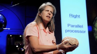
Jill Bolte Taylor
My stroke of insight, relevant talks.

VS Ramachandran
3 clues to understanding your brain.

Oliver Sacks
What hallucination reveals about our minds.
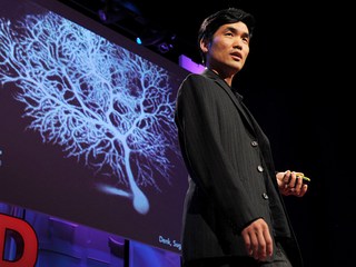
Sebastian Seung
I am my connectome.

Christopher deCharms
A look inside the brain in real time.

A light switch for neurons

Rebecca Saxe
How we read each other's minds.
- Biology Article
- Human Brain
The human brain controls nearly every aspect of the human body ranging from physiological functions to cognitive abilities. It functions by receiving and sending signals via neurons to different parts of the body. T he human brain, just like most other mammals, has the same basic structure, but it is better developed than any other mammalian brain.
Table of Contents
Brain Diagram
- Human Brain Parts
Read on to explore the human brain structure, diagram, parts of the human brain and the body functions controlled by the human brain.
Also Read: Placebo effect
Find out how some people live with just half a brain.

The Human Brain
On average, an adult brain weighs between 1.0 kg – 1.5 kg. It is mainly composed of neurons – the fundamental unit of the brain and nervous system. Recent estimates have suggested that the brain contains anywhere between 86 billion to 100 billion neurons.
The brain , along with the spinal cord , constitutes the central nervous system. It is responsible for thoughts, interpretation and origin of control for body movements.
Read More: Central Nervous System
The brain diagram given below highlights the different lobes of the human brain.

Where is the Brain located?
The brain is enclosed within the skull, which provides frontal, lateral and dorsal protection. The skull consists of 22 bones, 14 of which form the facial bones and the remaining 8 form the cranial bones. Anatomically, the brain is contained within the cranium and is surrounded by the cerebrospinal fluid.
The Cerebrospinal Fluid (CSF) is a fluid that circulates within the skull and spinal cord, filling up hollow spaces on the surface of the brain. Every day, the specialised ependymal cells produce around 500mL of cerebrospinal fluid.
The primary function of the CSF is to act as a buffer for the brain, cushioning mechanical shocks and dampening minor jolts. It also provides basic immunological protection to the brain.
Furthermore, CSF provides buoyancy for the brain. i.e., the brain is suspended in a layer of CSF, wherein, the weight of the brain is nearly negated. If the brain is not suspended in CSF, it would be impeded by its weight, consequently cutting off the blood supply in the lower half of the brain. It would lead to the death of neurons in the affected area.
Also Read : Neurons – Nerve Impulses
Parts of Human Brain
Following are the major parts of the human brain:
Forebrain – Largest part of the brain
It is the anterior part of the brain. The forebrain parts include:
- Hypothalamus
Forebrain Function: Controls the reproductive functions, body temperature, emotions, hunger and sleep.
Fact : The largest among the forebrain parts is the cerebrum. It is also the largest part of all vertebrate brains.
Midbrain : Smallest and central part of the brain
The midbrain consists of:
Read more: Tectum and Tegmentum
Hindbrain : The lower part of the brain
The hindbrain is composed of:
Hindbrain functions: The three regions of the hindbrain coordinates all processes necessary for survival. These induce breathing, heartbeat, sleep, wakefulness and motor learning.

Brain diagram highlighting various parts of the human brain
The cerebrum is the largest part of the brain. It consists of the cerebral cortex and other subcortical structures. It is composed of two cerebral hemispheres that are joined together by heavy, dense bands of fibre called the corpus callosum. The cerebrum is further divided into four sections or lobes:
Frontal lobe : It is associated with parts of speech, planning, reasoning, problem-solving and movements.
Parietal lobe : Help in movements, the perception of stimuli and orientation.
Occipital lobe : It is related to visual processing.
Temporal lobe : This region is related to perception and recognition of memory, auditory stimuli and speech.
The brain consists of two types of tissues: Grey matter and White matter .
- Grey matter mainly consists of various types of cells, which make up the bulk of the brain.
- White matter is primarily composed of axons, which connect various grey matter areas of the brain with each other.
The exterior portion of the cerebrum is called the cortex or the cerebral mantle. The cortex is extremely convoluted, due to which, it has a large surface area. The cerebrum also includes:
Sensory areas : To receive the messages.
Association areas : These areas integrate the incoming sensory information. It also forms a connection between sensory and motor areas.
Motor areas : This area is responsible for the action of the voluntary muscles.
Cerebrum Function
The cerebrum is responsible for thinking, intelligence, consciousness and memory. It is also responsible for interpreting touch, hearing and vision.
The thalamus is a small structure, located right above the brain stem responsible for relaying sensory information from the sense organs . It is also responsible for transmitting motor information for movement and coordination. Thalamus is found in the limbic system within the cerebrum. This limbic system is mainly responsible for the formation of new memories and storing past experiences.
The hypothalamus is a small and essential part of the brain, located precisely below the thalamus. It is considered the primary region of the brain, as it is involved in the following functions:
- Receives impulses
- Regulates body temperature
- Controls the mood and emotions
- Controls the sense of taste and smell
- Synthesises the body’s essential hormones
- Coordinates the messages from the autonomous nervous system
- Controls appetite, peristalsis, the rate of heartbeat, and blood pressure
- Forms an axis with the pituitary gland which is the main link between the nervous and the endocrine systems
Explore More: Hypothalamus
The tectum is a small portion of the brain, specifically the dorsal part of the midbrain. It serves as a relay centre for the sensory information from the ears to the cerebrum. It also controls the reflex movements of the head, eye and neck muscles. It provides a passage for the different neurons moving in and out of the cerebrum.
Tegmentum is a region within the brainstem. It is a complex structure with various components, which is mainly involved in body movements, sleep, arousal, attention, and different necessary reflexes. It forms the platform for the midbrain and connects with the thalamus, cerebral cortex and the spinal cord.
The cerebellum is the second largest part of the brain, located in the posterior portion of the medulla and pons. The cerebellum and cerebrum are separated by cerebellar tentorium and transverse fissure. Cortex is the outer surface of the cerebellum, and its parallel ridges are called the folia. Apart from this, the cerebellum has the cerebellar peduncles, cerebellar nuclei, anterior and posterior lobes. The cerebellum consists of two hemispheres, the outer grey cortex and the inner white medulla. It is mainly responsible for coordinating and maintaining the body balance during walking, running, riding, swimming, and precision control of the voluntary movements. The main functions of the cerebellum include:
- It senses equilibrium.
- Transfers information.
- Coordinates eye movement.
- It enables precision control of the voluntary body movements.
- Predicts the future position of the body during a particular movement.
- Both anterior and posterior lobes are concerned with the skeletal movements.
- The cerebellum is also essential for making fine adjustments to motor actions.
- Coordinates and maintains body balance and posture during walking, running, riding, swimming.
Explore: Differences between Cerebellum and Cerebrum
Medulla Oblongata
The medulla oblongata is a small structure present in the lowest region of the brain. It mainly controls the body’s autonomic functions such as heartbeat, breathing, and digestion. It plays a primary role in connecting the spinal cord, pons and the cerebral cortex. Also, it helps us in maintaining our posture and controlling our reflexes.
The pons is the primary structure of the brain stem present between the midbrain and medulla oblongata. It serves as a relay signals between the lower cerebellum, spinal cord, the midbrain, cerebrum and other higher parts of the brain. The main functions of the pons include:
- Controlling sleep cycles.
- Regulating the magnitude and frequency of the respiration.
- Transfers information between the cerebellum and motor cortex.
- Pons is also involved in sensations, such as the sense of taste, hearing and balance.
Further Reading: Interesting Facts about the Brain
To explore more about the human brain, parts of the human brain, its functions, brain diagram or any other related concept, please visit BYJU’S Biology
Frequently Asked Questions
1. explain how the nervous system is classified..
The nervous system in humans can be broadly classified into two types, namely, the peripheral nervous system and central nervous system
2. Describe the Central Nervous System.
The central nervous system primarily consists of the brain and the spinal cord. The system coordinates and controls various aspects of life, ranging from physical attributes (heartbeat, breathing) to mental capabilities (memory, intelligence).
3. Explain the Peripheral Nervous System.
The nerves and ganglia that are present outside the brain and spinal cord contribute to the peripheral nervous system. The primary role of the PNS is to connect the central nervous system to the rest of the body, such as the limbs, skin.
4. How is the brain protected in the body?
The brain is enclosed within the skull, where it is suspended in a layer of fluid called the cerebrospinal fluid. It protects the brain from minor mechanical shocks and jolts. Furthermore, it also serves minor immunological roles and provides the necessary nutrients required by the brain.
5. Briefly explain the parts of the brain.
Anatomically, the brain consists of the following parts:

Put your understanding of this concept to test by answering a few MCQs. Click ‘Start Quiz’ to begin!
Select the correct answer and click on the “Finish” button Check your score and answers at the end of the quiz
Visit BYJU’S for all Biology related queries and study materials
Your result is as below
Request OTP on Voice Call
Leave a Comment Cancel reply
Your Mobile number and Email id will not be published. Required fields are marked *
Post My Comment
Very helpful to study complex topics
this was a very helpful notes and questions for the preparation for class 10.
It’s very useful to understand some difficult topics
very useful notes for study. thank you
Fantastic study material
Super very interesting
Thank you byjus very helpful for me😃
Very amazing 😍 topic
Nice to study. so interesting
Nice content very useful for reading
Supper. Thank you so much. Really, this is a very good note. And it is very helpful for me.
Interesting and mind-blowing. Extraordinary👍💀🤯🤯
Thank you this site was very useful for my project 😊and I’ll look forward to taking all kinds of information from here
VERY NICE .THANK YOU .
Thank you, it was very useful. 🤓
very helpful I understand each and every thing thanks sir
good lesson
Nice concept, it is amazing and we can read it very easily😀😀😀😀😀😍😍😍😗😗😗
Very helpful!! Would surely recommend
very helpful notes thank you very much
VERY HELPFUL NOTES AND THANKS.
Byjus is very good, answering all questions
This is very interesting thank you so much
I AM VERY GRATEFUL TO HAVE BYJU’S IN THIS WORLD FOR HELPING ME WITH MY STUDIES
I like t way you teach but I think that you should also provide the whatsapp quary assistance also otherwise it is good keep it up👍👍👍👍👍👍👍👍
Thank you this site was very useful for my project 😊and I’ll look forward to taking all kinds of information from here 🥰🥰🥰🥰
- Share Share
Register with BYJU'S & Download Free PDFs
Register with byju's & watch live videos.

Human Brain Processes and Behavior Essay (Article)
Introduction.
The human brain is probably the most complex entity that is in existence or Nature. Memory, behavior, and the way the brain cells communicate are things that are sometimes difficult to contemplate, yet these processes take place every day.
Memory, retention, and recall are interconnected and often, influenced by the perspective of the person and associations they create for themselves. It is very relative and sometimes, false memories are created, either voluntarily or not. As emotions are always a part of human cognition, a certain predisposition and repetition of concepts lead a person to believe that something that was not present originally, was indeed there. As this area was little studied and several experiments have yielded data that cannot be heavily relied on, it stays the subject of research. The assumption is that in a specific situation the way the information is stored in the long term memory and the way it is later recalled is influenced by the person’s emotional state and their personal views, opinions, and perspectives. Stress, both positive and negative, has a great effect on the precision of recollection of events and the creation of false memories.
Traumatic experiences are remembered very vividly and emotions add certain details that might not have been present in reality. This is a valid point because a recollection of an event has several cognitive stimuli, and a person feels an individual and emotional connection to what is taking place. In the case with a simple word remembering, people do not have exposure to the situation or the emotional meaning of the words. For example, the word “betrayal” could mean one thing to someone who knows about it hypothetically, in comparison to someone who has lived through it a long time ago, and it could be even more meaningful to a person who has experienced it recently. The brain very much works on associations, for better storage and recall of information. A connection between two events or concepts, uniting them according to their similarities or specific differences, creates a link that makes it easier for the brain to remember (Hogenboom, 2013).
Psychology has long tried to pinpoint the definitive and singular reasons for human behaviors and attitudes. There are very many common variables, but each individual is unique in the end. Mimicking others is one of the links in an enormous chain of individuality that makes up each person. It is very true that the proximity of individuals heightens the sensitive nature of interaction within the human world. It is clear that people are very much connected to the family, relatives, friends, and partners and so, they gain great knowledge and illustration of what these people want in their life. This gives a person a closer look and comparison in regards to their own personality and wants. A suggestion is made that there is a possibility of the person who is most admired becoming the “obstacle” for the observer. This suggests a negative nature of closeness and relation between people.
But, an important point is that humans are social beings, and cooperation, together with mutual dependence and help are inevitable. There are many examples of people being more successful through positive competition and unity of interests where one person does not let greed and egoism to overtake their existence. It becomes clear that people will always compare themselves to their environment and others, which will lead to new efforts and imagination of each individual and collective humanity. Another confirmation that mimicry and competition is an important and needed part of the world is that it is present throughout the animal kingdom, where cruelty and violence should not be necessarily labeled as greedy and selfish. It is interesting that lower animals and humans have been known to mimic each other in behaviors and instincts. This shows that the repetition of seen behaviors is one of the simplest forms of action evident in all of nature (Thomas, 2013).
One of the most interesting discoveries made was about the communication between brain cells, axons, through their synapses. It was thought that there is a physical connection between each axon that was needed for the transfer of information. In reality, there is a gap between the end of the axon, its tip, and the dendrites of the next axon. Chemicals are being released by one axon and they float to the receptors of the next axon. The chemicals that are produced, neurotransmitters, are a sort of boats that carry their own information. Those chemicals that were not absorbed by the receiving axon, return to the source, the sending axon and are recycled.
The amount of repetitions of the chemicals being sent determines the strength of the signal. If a person wants something in a minor way, the signal is sent once or twice, and the receiving axon does not respond in a major way. But, in case the signal is repeated constantly, the axon is bombarded with chemicals, thus causing a conscious response from a person. This is what determines the cognition of a person. It is still unclear how a person’s mind or brain can hold so much information. It has been supposed that a person has unlimited potential. An amazing fact is that such a small entity as a cell can hold so much information and have significant energy to send impulses that affect the whole body. All the systems and different parts of the brain are connected, and studies have shown that trauma to a certain area will be compensated by another part. There are also cases when the loss of a connection between the brain divisions has led to the loss of speech, cognition, memory, and senses (Axon, 2013).
Even though people understand more about the human brain and behavior, there is still a lot to learn, which leaves the human brain one of the most mysterious and little-studied entities.
Axon. (2013). Web.
Hogenboom, M. (2013). Why does the human brain create false memories? Web.
Thomas, J. (2013). Mimicking Behavior May Help Form Social Bonds. Web.
- Chicago (A-D)
- Chicago (N-B)
IvyPanda. (2020, October 10). Human Brain Processes and Behavior. https://ivypanda.com/essays/human-brain-processes-and-behavior/
"Human Brain Processes and Behavior." IvyPanda , 10 Oct. 2020, ivypanda.com/essays/human-brain-processes-and-behavior/.
IvyPanda . (2020) 'Human Brain Processes and Behavior'. 10 October.
IvyPanda . 2020. "Human Brain Processes and Behavior." October 10, 2020. https://ivypanda.com/essays/human-brain-processes-and-behavior/.
1. IvyPanda . "Human Brain Processes and Behavior." October 10, 2020. https://ivypanda.com/essays/human-brain-processes-and-behavior/.
Bibliography
IvyPanda . "Human Brain Processes and Behavior." October 10, 2020. https://ivypanda.com/essays/human-brain-processes-and-behavior/.
- Peripheral Nervous System and Its Development
- Neurological System Disorder of Falling Asleep
- Cell Membrane Modeling and Cell Membrane Theory
- Thought Processes and Perception Influences
- Working Memory Training and Its Controversies
- False Memories in Patients with Depression
- Meditation's Effects on Inner Word and Perception
- Cognition and Decision-Making in Kahneman's Ideas
Home — Essay Samples — Nursing & Health — Anatomy & Physiology — Human Brain
Essays About Human Brain
Brief description of human brain.
The human brain is the most complex organ in the body, responsible for controlling our thoughts, emotions, and actions. It plays a crucial role in our everyday functioning, from simple tasks like breathing and walking to complex activities like problem-solving and decision-making.
Importance of Writing Essays on ... Read More Brief Description of Human Brain
Importance of writing essays on this topic.
Essays on the human brain are essential for understanding the intricate workings of the brain and its impact on human behavior, cognition, and mental health. They also provide a platform for exploring the latest research and advancements in neuroscience, psychology, and related fields.
Tips on Choosing a Good Topic
- Focus on current issues and debates in neuroscience and psychology.
- Explore the relationship between the brain and behavior in specific populations, such as children, the elderly, or individuals with neurological disorders.
- Consider interdisciplinary approaches that integrate neuroscience with other fields, such as philosophy, ethics, or artificial intelligence.
Essay Topics
- The impact of stress on the brain and mental health.
- The role of neurotransmitters in regulating mood and behavior.
- Exploring the link between brain injuries and cognitive impairment.
- The ethical implications of brain-computer interfaces.
- The influence of genetics on brain development and functioning.
- The neurobiology of addiction and substance abuse.
- The neurological basis of memory and learning.
- The effects of meditation and mindfulness on brain function.
- The relationship between sleep patterns and brain health.
- The future of artificial intelligence and its impact on the human brain.
Concluding Thought
Writing essays on the human brain offers a unique opportunity to delve into the complexities of the mind and gain a deeper understanding of what makes us human. By exploring diverse topics within this field, we can contribute to the collective knowledge and foster critical thinking about the brain and its profound influence on our lives.
Brain - The Most Important Organ
Brain as the main part of a body, made-to-order essay as fast as you need it.
Each essay is customized to cater to your unique preferences
+ experts online
Anatomy of The Brain
The left-brain-right-brain theory of personality, the role of brain in behavior, three different types of human memory, let us write you an essay from scratch.
- 450+ experts on 30 subjects ready to help
- Custom essay delivered in as few as 3 hours
Computer as a Way to Expand The Brain
Review on working memory, how cell phone addiction affects the brain, neuroscience & applied cognitive sciences, get a personalized essay in under 3 hours.
Expert-written essays crafted with your exact needs in mind
Critically Assess How General Anaesthetics Cause Loss of Consciousness
A brain aneurysm, musical memory and alzheimer's disease, headache as the symbol of brain cancer, overview of the aging brain issue, video games can be beneficial for our health and brain, improved brain tumor detection, brainstem: review of human brain, artificial intelligence can possibly replace human brain, biological basis of addiction, overview of gliomas and its histopathology, neurological disorder of epilepsy: diagnose and treatment, the brain: correlation, unity, and damage, the mechanism of the human memory, the creation of our memories, how parkinson’s disease affects your body, my interest to obtain knowledge on the threats and opportunities posed by synthetic biology on security and safety, hermann grid's review, the formation of habits in the brain, brain aneurysm: causes, symptoms, treatment, relevant topics.
- Human Physiology
- Cardiovascular System
- Human Anatomy
- Homeostasis
- Respiratory System
- Digestive System
By clicking “Check Writers’ Offers”, you agree to our terms of service and privacy policy . We’ll occasionally send you promo and account related email
No need to pay just yet!
We use cookies to personalyze your web-site experience. By continuing we’ll assume you board with our cookie policy .
- Instructions Followed To The Letter
- Deadlines Met At Every Stage
- Unique And Plagiarism Free

How does the brain think?

Professor of Psychology, Auburn University
Disclosure statement
Dr. Jennifer L. Robinson is a professor in the Department of Psychological Sciences at Auburn University. She also serves as a paid consultant for VDF FutureCeuticals, Inc. and has received funding from the company to carry out clinical trials.
Auburn University provides funding as a member of The Conversation US.
View all partners

Curious Kids is a series for children of all ages. If you have a question you’d like an expert to answer, send it to [email protected] .
How does the brain think? – Tom, age 16, San Diego, California
Have you ever wondered how your brain creates thoughts or why something randomly popped into your head? It may seem like magic – but actually the brain is like a supercomputer inside your head that helps you think, learn and make decisions .
Imagine your brain as a busy city with lots of streets and buildings. Each part of the brain has a specific job to do, just like certain areas of a city or certain buildings serve different purposes. When you have a thought, it’s like a message traveling through the city, passing from one area to another.
As a professor of psychology and neuroscience , I have studied the brain for almost 20 years. Neurologists , neuroscientists and neurosurgeons work every day to understand the brain better. And there’s still a lot to learn.
Practice and repetition create skills
The neuron is a key player in the brain – these are tiny cells that send and receive signals and messages so they can communicate with each other.
Your brain has somewhere between 80 billion and 100 billion neurons . Neurons tend to group together to form neural tracts , which would be like the streets and highways in the city analogy. When you have a thought, neurons in your brain fire up and create electrical impulses. These impulses tend to travel along similar pathways and release tiny chemicals called neurotransmitters along the way.
These neurotransmitters are like the construction crew that builds the roads, making it easier for the messages to be delivered. You can imagine it as a dirt road, but as more traffic – that is, neuron signals – travel the dirt road, the road gets upgraded to a paved street. If the traffic continues, it gets upgraded to a highway.
As you learn new things and experience the world around you, these connections grow stronger . For example, when you are learning to ride a bike, you may be unsteady and find it hard to coordinate all of the different muscles along with your ability to balance. But the more you practice, the more the neurons controlling your muscles and your ability to balance fire together, which makes it much easier as you practice. Neurons are wiring together and forming neural networks.
That’s why practice and repetition are important for improving your skills, whether playing the piano or learning a language. Neural networks are created and then strengthened the more times they communicate together. Scientists have a saying in this field: “Neurons that fire together wire together.” Certain thinking or behavior patterns can be chalked up to this kind of repeated synchronized activity.
Developing creativity
You are conscious of only a very small portion of the information your brain takes in. It is constantly receiving input from your senses – sights, sounds, tastes, smells and touch. When you see a cute puppy or hear your favorite song, your senses send signals to the brain, triggering a chain reaction of thoughts and emotions.
The brain also stores memories , which are like files in a computer that you can access whenever you need them. Memories help shape your thoughts and influence how you see the world.
If you remember a fun day at the beach, it might make you feel happy and relaxed. If you smell an apple pie, it may remind you of your grandma’s baking. These thoughts are triggered because these pleasant associations have been formed in your brain, and through repetition, strengthened over time.
Creativity is another superpower of the brain. When you let your imagination run wild, your brain can come up with new ideas, stories and inventions . Artists, writers and scientists all use their creative brains to explore new possibilities and solve problems.
Have you ever experienced a “eureka” moment when a brilliant idea pops into your head out of nowhere? That’s your brain’s way of connecting the dots and coming up with a solution.
Keeping your brain healthy
Most scientists agree that sleep is really important for your brain to process information from the day and to allow it to rest and form new connections. A lot of people find that they have new ideas or thoughts after a good night’s sleep. The opposite is true, too – without enough sleep, you may feel like you can’t think straight.
Along with enough sleep, eat healthy foods and exercise . Just like a car needs fuel to run smoothly, your brain needs nutrients and oxygen to function at its best and to boost your thinking power.
Activities that challenge you are also great: reading , doing puzzles, playing music, making art, doing math , writing essays and book reports and journaling . Positive thinking also helps. Keep in mind that whatever you are consuming – what you’re eating or what you’re watching, listening to or reading – has the power to influence your brain.
Conversely, smoking cigarettes , vaping , drinking alcohol and using drugs kills brain cells. So might head injuries that can occur when playing sports such as football, soccer and bicycling – but wearing a helmet can make a big difference .
The brain is a fascinating organ that works tirelessly to create thoughts, memories and ideas. As technology continues to improve, scientists will learn more and more about how biological processes give rise to our conscious experiences. The challenges of learning about the brain are like a neuroscientific moonshot – we have a long way to go before we completely understand how it works.
Hello, curious kids! Do you have a question you’d like an expert to answer? Ask an adult to send your question to [email protected] . Please tell us your name, age and the city where you live.
And since curiosity has no age limit – adults, let us know what you’re wondering, too. We won’t be able to answer every question, but we will do our best.
- Neuroscience
- Curious Kids
- Curious Kids US
Events and Communications Coordinator
Assistant Editor - 1 year cadetship
Executive Dean, Faculty of Health
Lecturer/Senior Lecturer, Earth System Science (School of Science)
Sydney Horizon Educators (Identified)
- Alzheimer's disease & dementia
- Arthritis & Rheumatism
- Attention deficit disorders
- Autism spectrum disorders
- Biomedical technology
- Diseases, Conditions, Syndromes
- Endocrinology & Metabolism
- Gastroenterology
- Gerontology & Geriatrics
- Health informatics
- Inflammatory disorders
- Medical economics
- Medical research
- Medications
- Neuroscience
- Obstetrics & gynaecology
- Oncology & Cancer
- Ophthalmology
- Overweight & Obesity
- Parkinson's & Movement disorders
- Psychology & Psychiatry
- Radiology & Imaging
- Sleep disorders
- Sports medicine & Kinesiology
- Vaccination
- Breast cancer
- Cardiovascular disease
- Chronic obstructive pulmonary disease
- Colon cancer
- Coronary artery disease
- Heart attack
- Heart disease
- High blood pressure
- Kidney disease
- Lung cancer
- Multiple sclerosis
- Myocardial infarction
- Ovarian cancer
- Post traumatic stress disorder
- Rheumatoid arthritis
- Schizophrenia
- Skin cancer
- Type 2 diabetes
- Full List »
share this!
May 9, 2024
This article has been reviewed according to Science X's editorial process and policies . Editors have highlighted the following attributes while ensuring the content's credibility:
fact-checked
peer-reviewed publication
trusted source
Human brain map contains never-before-seen details of structure
by Harvard University

A cubic millimeter of brain tissue may not sound like much. But considering that tiny square contains 57,000 cells, 230 millimeters of blood vessels, and 150 million synapses, all amounting to 1,400 terabytes of data, Harvard and Google researchers have just accomplished something enormous.
A Harvard team led by Jeff Lichtman, the Jeremy R. Knowles Professor of Molecular and Cellular Biology and newly appointed dean of science, has co-created with Google researchers the largest synaptic-resolution, 3D reconstruction of a piece of human brain to date, showing in vivid detail each cell and its web of neural connections in a piece of human temporal cortex about half the size of a rice grain.
The feat, published in Science , is the latest in a nearly 10-year collaboration with scientists at Google Research, who combine Lichtman's electron microscopy imaging with AI algorithms to color-code and reconstruct the extremely complex wiring of mammal brains. The paper's three co-first authors are former Harvard postdoctoral researcher Alexander Shapson-Coe; Michał Januszewski of Google Research, and Harvard postdoctoral researcher Daniel Berger.
The collaboration's ultimate goal is to create a high-resolution map of a whole mouse brain's neural wiring, which would entail about 1,000 times the amount of data they just produced from the 1-cubic-millimeter fragment of human cortex.
"The word 'fragment' is ironic," Lichtman said. "A terabyte is, for most people, gigantic, yet a fragment of a human brain—just a miniscule, teeny-weeny little bit of human brain—is still thousands of terabytes."
The latest map in Science contains never-before-seen details of brain structure, including a rare but powerful set of axons connected by up to 50 synapses. The team also noted oddities in the tissue, such as a small number of axons that formed extensive whorls. Since their sample was taken from a patient with epilepsy, they're unsure if such unusual formations are pathological or simply rare.
Lichtman's field is "connectomics," which, analogous to genomics, seeks to create comprehensive catalogs of brain structure, down to individual cells and wiring. Such completed maps would light the way toward new insights into brain function and disease, about which scientists still know very little.
Google's state-of-the-art AI algorithms allow for reconstruction and mapping of brain tissue in three dimensions. The team has also developed a suite of publicly available tools researchers can use to examine and annotate the connectome.
"Given the enormous investment put into this project, it was important to present the results in a way that anybody else can now go and benefit from them," said Google Research collaborator Viren Jain.
Next the team will tackle the mouse hippocampal formation, which is important to neuroscience for its role in memory and neurological disease.
Explore further
Feedback to editors

Study links organization of neurotypical brains to genes involved in autism and schizophrenia
17 minutes ago

Study traces an infectious language epidemic
8 hours ago

Visual experiences unique to early infancy provide building blocks of human vision, study finds
18 hours ago

Study points to personalized treatment opportunities for glioblastoma
19 hours ago

Research team introduces new tool to boost battle against childhood undernutrition

How herpes hijacks a ride into cells
20 hours ago

How the brain is flexible enough for a complex world, without being thrown into chaos

Researchers create AI model to understand how brain activity relates to illness

Study reveals need to review temperature control measures in hospitals to manage Legionella

'What was that?' How brains convert sounds to actions
21 hours ago
Related Stories

Google teams up with Harvard to map small bit of human cortex
Jun 8, 2021

Connecting lab-grown brain cells provides insight into how our own brains work
Apr 10, 2024

Unlocking mysteries of octopus cognition: Paving the way for memory research
Aug 16, 2023

Researchers develop technology to tabulate and characterize every cell in the human brain
Oct 12, 2023

Deep inside the brain: Mapping the dense neural networks in the cerebral cortex
Oct 25, 2019

Why a tiny worm's brain development could shed light on human thinking
Aug 4, 2021
Recommended for you

Psychological therapy shows promise in improving quality of life for people living with motor neuron disease
22 hours ago

Using MRI, engineers have found a way to detect light deep in the brain
23 hours ago
Let us know if there is a problem with our content
Use this form if you have come across a typo, inaccuracy or would like to send an edit request for the content on this page. For general inquiries, please use our contact form . For general feedback, use the public comments section below (please adhere to guidelines ).
Please select the most appropriate category to facilitate processing of your request
Thank you for taking time to provide your feedback to the editors.
Your feedback is important to us. However, we do not guarantee individual replies due to the high volume of messages.
E-mail the story
Your email address is used only to let the recipient know who sent the email. Neither your address nor the recipient's address will be used for any other purpose. The information you enter will appear in your e-mail message and is not retained by Medical Xpress in any form.
Newsletter sign up
Get weekly and/or daily updates delivered to your inbox. You can unsubscribe at any time and we'll never share your details to third parties.
More information Privacy policy
Donate and enjoy an ad-free experience
We keep our content available to everyone. Consider supporting Science X's mission by getting a premium account.
E-mail newsletter
See the most detailed map of human brain matter ever created
Explore the tiny 'pizza slice' down to the neuron.
By Lauren Leffer | Published May 9, 2024 2:00 PM EDT

A cubic millimeter is, by all accounts, tiny. It’s barely noticeable–a speck or fleck or crumb. But look closely enough and you can uncover an entire world inside a particle of material. A team of neuroscientists and engineers, aided by machine learning tools, have charted a cubic millimeter volume of the human brain at nanoscale resolution, tracing every neuron, synapse, blood vessel, and supporting cell within the fragment and reconstructing a 3D model of the tissue. Though it represents just one-millionth of the total brain volume, it is the most detailed map of a piece of human brain matter ever created. It could spur a wave of scientific discovery about neurological disorders, brain structure, and the origins of our behavior.
“In one respect, our data set is miniscule,” Jeff Lichtman , co-senior study researcher and a neuroscientist and professor of molecular and cellular biology at Harvard University, tells PopSci . “But it doesn’t feel small because when you get in it, you see it’s like a gigantic forest. It’s a very tiny forest, but it’s a very, very, very complicated forest,” he adds.

All that complexity is on display in a study documenting the construction of this bit of comprehensive brain map or “connectome,” published May 9 in the journal Science . The first connectome was of a nematode brain, completed in 1986 . Since then, neuroscientists have continued to plot out increasingly large and complicated brains–including those of fruit flies, maggots, a tadpole, and an earthworm. Yet human brains pose a unique mapping challenge in their intricacy and inaccessibility. The new, partial human connectome is available online for anyone to explore.
“Not only is this an impressive technological feat, this is a tool and a resource that is really aimed at sharing with the world and getting all of this scientific information out there,” Tim Mosca , a neuroscientist at Thomas Jefferson University who was uninvolved in the new work, tells PopSci . “This group has done an amazing job designing all of the new tools and the pipelines to make this available to anyone who wants to look at it, wants to think about it, wants to use this in their research.”

Serving up brain pizza
The study sample was collected over a decade ago from an anonymous patient undergoing epilepsy surgery. The surgeon removed a small piece of the temporal lobe to access and treat an underlying lesion, quickly preserved the tissue, and later shared it with scientists. Though the total volume of the fragment is about 1 cubic millimeter, it is not cube shaped. Instead, “it’s like a thick piece of pizza–but it’s not that thick,” says Lichtman. This blunt, triangular chunk, longer than it is wide, enabled the researchers to capture a bit of all six layers of the 3mm thick cerebral cortex.

The first step to mapping the brain pizza was to slice it into 5,019 individual cross sections (each 30 nanometers thin) mounted on tape using a specially designed machine that cuts with a diamond knife. From there, the researchers spent a full year carefully imaging each slice via electron microscope. Then, they digitally aligned and stitched together the slices and used multiple machine learning tools to fill out the 3-D form and label and color each component.
The segment’s neuron density is 16,000 neurons per cubic millimeter– about one-third lower than a previous density estimate of the same brain section and 10 times less dense than the corresponding section of a mouse’s brain, per the study. Glial cells, the connective glue that keeps brain tissue together, outnumber neurons in the fragment by a two to one ratio.
Neural explorers
The physical size of the brain fragment may be teeny, but the level of detail means the data captured by the mapping effort is massive. The reconstructed segment is 1.4 petabytes in digital size, or 1,400 terabytes (equivalent to the storage capacity of about 2,800 average laptops). Within that, there is lots to potentially discover: individual neural circuits, previously unobserved cellular ratios and shapes, the makeup of each cortical layer, and more.
“It’s like being an explorer that lands on a new island,” says Lichtman. “You keep looking around and you’re just going to keep finding new things.”
Already, Lichtman and his many co-researchers have made some interesting observations. Amid the ~150 million synapses they mapped, they found a rare type of particularly strong connection. In the vast majority (96.5%) of cases, axons–the outgoing transmission line of neurons– formed one connection with a target cell. Some (about 3%) made 2 connections. But less than .01% forged more than four synapses, including some axons and target cells that were connected at over 50 points.

“We’ve always had a theory that there would be super connections, if you will, amongst certain cells,” says Mosca. “But it’s something we’ve never had the resolution to prove…Now we know that it exists and we can go after the question of what it does.” Lichtman’s current hypothesis is that these extra-fortified connections are the sort of hyper-fast pathways that enable “automatic use of the brain” for well-established, learned actions.
Another new observation: many dendrites (the branching extensions of neurons that generally receive inputs) seem to mirror each other–pointing symmetrically in one of just two directional arrangements out of infinite three-dimensional possibilities. “We’d never seen anything like that [before],” says Lichtman. “Why are they doing that? We don’t know… [it is] a complete mystery.”

The scientists further found a new type of unexplained structure that they’ve named an “axon whorl,” wherein long axon cables appear to tangle around themselves. Though it wasn’t every neuron, some axons contained multiple knots, says Viren Jain , co-senior study author and a senior staff scientist at Google where he leads the company’s Connectomics research team. Again, the function and cause of these whorls is unknown. “We were not expecting to find such a structure. It’s very peculiar… like a big jumble of wiring that sort of contravenes the purpose of a wire to begin with, which is to go places and contact other things.”

These three findings are likely just the tip of the iceberg. “The data set is so large that one human being or lab group can not explore it [all], but a bunch of human beings can,” Lichtman says. Because of the open nature of the project, more than 200 papers have cited the brain reconstruction since it was first released as a pre-print, Jain points out.
In addition to being a large, fundamental advance in science, discoveries resulting from this partial connectome could eventually help us better understand and treat brain diseases. “The ability to measure neural wiring of human brains in such detail opens up exciting opportunities for advancing human health,” says Andrew Leifer , a physicist and neuroscientist at Princeton University who wasn’t involved in the project. “One could imagine comparing different brains to understand how brain wiring changes when a healthy brain suffers from a disease or falls into dysfunction,” he adds.
Pushing into future frontiers
But though there is lots to be discovered, there are also limits. The automated machine learning methods which were key to enabling such a large-scale endeavor carry a margin of error that requires human oversight to correct. Editing will be an ongoing project, and is a community science effort anyone who wants to can apply to participate in .
The sample is also only one small piece of one person’s brain. There is much that can’t yet be inferred about human brains generally or other brain regions beyond the temporal lobe based on this single fragment without more samples and maps for comparison, notes Lichtman.
And, perhaps most critically, the brain segment came from someone undergoing surgery for epilepsy–it may not represent a “normal” brain and there’s no way to know for sure unless and until we have more bits to assess, say Jain and Lichtman. “But we are planning many follow-ups to this,” Jain adds.

The team has ambitions to construct multiple partial connectomes representing additional human brain samples. They are also working on zebrafish connectome, and are planning to tackle increasingly large segments of the mouse brain. Mammalian brains share many similarities, so a complete mouse connectome could offer new insights into our own brain as well as the evolution of brains across animals, Lichtman says.
At the moment, with currently available technologies (and the ethical implications), a complete connectome of the human brian is “a bridge too far,” says Lichtman. “Literally, we’re a million times away from that,” says Jain. But through this study, the scientists have taken an early (if miniscule) step in that direction, and even the smallest peephole can be a portal into a whole universe of knowledge.
“I would love people to think about this the same way they think about the Hubble or James Webb telescope,” says Lichtman. “We’re peering into an unknown domain, and one that is much more relevant to us than distant outer space. It’s this inner space that each of us have on our shoulders that we use, but know almost nothing about.”
Like science, tech, and DIY projects?
Sign up to receive Popular Science's emails and get the highlights.
Scientists Imaged and Mapped a Tiny Piece of Human Brain. Here’s What They Found
With the help of an artificial intelligence algorithm, the researchers produced 1.4 million gigabytes of data from a cubic millimeter of brain tissue
/https://tf-cmsv2-smithsonianmag-media.s3.amazonaws.com/accounts/headshot/Will-Sullivan-photo.png "parts of human brain essay")
Will Sullivan
Daily Correspondent
:focal(1664x1093:1665x1094)/https://tf-cmsv2-smithsonianmag-media.s3.amazonaws.com/filer_public/c7/ba/c7baacef-cb16-4351-bd03-27bd56c1b461/connectomics2024-1-excitatoryneuronsoriginal.png "parts of human brain essay")
Researchers have made a digital map showing a tiny chunk of a human brain in unprecedented detail.
Based on a brain tissue sample that had been surgically removed from a person, the map represents a cubic millimeter of brain—an area about half the size of a grain of rice. But even that tiny segment is overflowing with 1.4 million gigabytes of information—containing about 57,000 cells, 230 millimeters of blood vessels and 150 million synapses, the connections between neurons.
The researchers published their findings in the journal Science on Friday. They have made the data set freely available online and provided tools for analyzing and proofreading it.
“In one respect, our data set is miniscule,” Jeff Lichtman , a co-author of the study and biologist at Harvard University, tells Popular Science ’s Lauren Leffer. “But it doesn’t feel small, because when you get in it, you see it’s like a gigantic forest.”
“It’s a little bit humbling,” Viren Jain , a co-author of the study and neuroscientist at Google, says to Nature News ’ Carissa Wong. “How are we ever going to really come to terms with all this complexity?”
The first such map of a brain was made in 1986, when researchers cataloged the 302 neurons of a roundworm, Jain writes in a Google blog post about the new research. In 2020, the Google team mapped 25,000 neurons in part of a fruit fly brain. They’ve also been involved in mapping parts of the brains of the zebra finch (a small bird) and zebrafish larvae.
For the new paper, the researchers turned to a piece of brain tissue from a person with epilepsy. To access a lesion in the patient’s hippocampus, a surgeon had removed the tissue from their left anterior temporal lobe , a part of the brain thought to play a role in our memory of objects, people, words and facts.
Scientists then used an electron microscope to image more than 5,000 slices of the tissue, each of which was only around 30 nanometers thick. The process took close to 11 months. Artificial intelligence algorithms then reconstructed the cells and their connections in 3D.
“The aim was to get a high resolution view of this most mysterious piece of biology that each of us carries around on our shoulders,” Lichtman tells the Guardian ’s Ian Sample. “The reason we haven’t done it before is that it is damn challenging. It really was enormously hard to do this.”
“It’s probably the most computer-intensive work in all of neuroscience,” Michael Hawrylycz , a computational neuroscientist at the Allen Institute for Brain Science who did not contribute to the findings, tells MIT Technology Review ’s Cassandra Willyard. “There is a Herculean amount of work involved.”
The data has already yielded some unexpected findings for the researchers. “There were just so many things in it that were incompatible with what you would read in a textbook,” Lichtman says to MIT Technology Review .
For instance, they found some rare sites where neurons were connected by more than 50 synapses. This is incredibly uncommon—more than 96 percent of connections between neurons have just one synapse, and more than 99 percent have three or fewer synapses, per Google’s blog post.
These strong connections might show “what learning looks like in the brain,” Lichtman hypothesizes to the Guardian , representing behaviors so well-practiced that they require almost no thought at all, such as moving your foot from the accelerator to the brake when stopping a car.
The team also found cases of axons, the tendrils on neurons that transmit messages, wrapping themselves into knots—but they don’t know why. “Nobody had seen anything like this before,” Jain tells Nature News .
While the tissue sample didn’t have any evidence of disease, the researchers can’t be certain whether these surprises are in some way related to the person’s epilepsy or treatments they were receiving. Studying other tissue samples could give them a clearer idea.
Next, the researchers would like to map the hippocampus of a mouse. But in the meantime, the human data they have so far will allow other researchers the chance to learn more about the human brain.
“Not only is this an impressive technological feat, this is a tool and a resource that is really aimed at sharing with the world and getting all of this scientific information out there,” Tim Mosca , a neuroscientist at Thomas Jefferson University who did not contribute to the findings, tells Popular Science . “This group has done an amazing job designing all of the new tools and the pipelines to make this available to anyone who wants to look at it, wants to think about it, wants to use this in their research.”
Get the latest stories in your inbox every weekday.
/https://tf-cmsv2-smithsonianmag-media.s3.amazonaws.com/accounts/headshot/Will-Sullivan-photo.png "parts of human brain essay")
Will Sullivan | | READ MORE
Will Sullivan is a science writer based in Washington, D.C. His work has appeared in Inside Science and NOVA Next .
IMAGES
VIDEO
COMMENTS
The human brain is a complex organ, made up of several distinct parts, each responsible for different functions. The cerebrum, the largest part, is responsible for sensory interpretation, thought processing, and voluntary muscle activity. Beneath it is the cerebellum, which controls balance and coordination. The brainstem connects the brain to the spinal cord and oversees automatic processes ...
Summary. The brain connects to the spine and is part of the central nervous system (CNS). The various parts of the brain are responsible for personality, movement, breathing, and other crucial ...
The cerebellum ("little brain") is a fist-sized portion of the brain located at the back of the head, below the temporal and occipital lobes and above the brainstem. Like the cerebral cortex, it has two hemispheres. The outer portion contains neurons, and the inner area communicates with the cerebral cortex.
The three main parts of the brain are the cerebrum, cerebellum, and brainstem. 1. Cerebrum. Location: The cerebellum occupies the upper part of the cranial cavity and is the largest part of the human brain. Functions: It's responsible for higher brain functions, including thought, action, emotion, and interpretation of sensory data.
This area houses the brain's "gray matter," and is considered the "seat" of human consciousness. Higher brain functions such as thinking, reasoning, planning, emotion, memory, the processing of sensory information and speech all happen in the cerebral cortex. In other words, the cerebral cortex is what sets humans apart from other species.
brain, the mass of nerve tissue in the anterior end of an organism. The brain integrates sensory information and directs motor responses; in higher vertebrates it is also the centre of learning.The human brain weighs approximately 1.4 kg (3 pounds) and is made up of billions of cells called neurons.Junctions between neurons, known as synapses, enable electrical and chemical messages to be ...
The human brain is a 3-pound (1.4-kilogram) mass of jelly-like fats and tissues—yet it's the most complex of all known living structures. The brain is extremely sensitive and delicate, and so it ...
The human brain is perhaps the most complex of all biological systems, with the mature brain composed of more than 100 billion information-processing cells called neurons.[1] The brain is an organ composed of nervous tissue that commands task-evoked responses, movement, senses, emotions, language, communication, thinking, and memory. The three main parts of the human brain are the cerebrum ...
The brain is the most complex organ in the human body. It produces our every thought, action, memory, feeling and experience of the world. This jelly-like mass of tissue, weighing in at around 1.4 ...
Everything you ever wanted to know about the brain: here are answers to the most popular questions about the human brain. Explore the wonders of your grey matter, the organ that makes you you.
The cerebellum makes up approximately 10% of the brain's total size, but it accounts for more than 50% of the total number of neurons located in the entire brain. The cerebellum is comprised of small lobes and serves several functions. It receives information from the inner ear's balance system, sensory nerves, and auditory and visual systems.
The brain works like a big computer. It processes information that it receives from the senses and body, and sends messages back to the body. But the brain can do much more than a machine can: We think and experience emotions with our brain, and it is the root of human intelligence. The human brain is roughly the size of two clenched fists and weighs about 1.5 kilograms. From the outside it ...
Introductory essay. Written by the educators who created Mapping and Manipulating the Brain, a brief look at the key facts, tough questions and big ideas in their field. Begin this TED Study with a fascinating read that gives context and clarity to the material. Here is this mass of jelly, three-pound mass of jelly you can hold in the palm of ...
The cerebrum is another part of the human brain located in the uppermost section. Its main role in the body is to coordinate the function and processing of the sensory functions. These include the main body senses, such as vision, hearing, and touch necessary for the body's normal functioning. This function aids in controlling body movement ...
The brain and its function. comprise a central nervous system. A brainstem, which in part relays information between the peripheral nerves and spinal. cord to the upper parts of the brain consists ...
The human brain controls nearly every aspect of the human body ranging from physiological functions to cognitive abilities. It functions by receiving and sending signals via neurons to different parts of the body. The human brain, just like most other mammals, has the same basic structure, but it is better developed than any other mammalian brain.
The cerebrum, also known as the cerebral cortex, is the biggest portion of the human brain, linked with higher brain functions such as action and thought. The cerebral cortex is partitioned into four segments, referred to as lobes: the frontal lobe, parietal lobe, occipital lobe, and temporal lobe. Below is a diagram of the cerebrum:
Essay on Human Brain: Structure and Function. The nervous system of man and other group of vertebrates is divided into three main parts: 1. Central nervous system (CNS) comprising brain and spinal cord. 2. Peripheral nervous system (PNS) consisting of cranial and spinal nerves. 3.
Introduction. The human brain is probably the most complex entity that is in existence or Nature. Memory, behavior, and the way the brain cells communicate are things that are sometimes difficult to contemplate, yet these processes take place every day. We will write a custom essay on your topic. 809 writers online.
Essay about the human brain. In this paper one will learn the different parts of the brain and their functions. Although the brain isn't the largest organ of the human body it is the most complex and controlling organ. It is amazing how complicated the brain is. The brain controls every action within and out of your body.
Essays on the human brain are essential for understanding the intricate workings of the brain and its impact on human behavior, cognition, and mental health. ... Working memory is a part of the human memory system with a limited capacity. Working Memory's job is holding temporarily information for processing. Working memory refers to ...
In the brain, nerve centers corresponding. to reflex reflex es such as [ 6-7, 12] are located: breathing, ingestion, suction, cough, sneezing, blinking, et c. 3.2 Emotions and limbic system. The ...
Each part of the brain has a specific job to do, just like certain areas of a city or certain buildings serve different purposes. ... doing math, writing essays and book reports and journaling ...
Human Brain One of the most complex organs in the universe, the human brain, continues to be a scientific mystery. In vertebrate and most invertebrate animals, the brain is the central aspect of the nervous system. The brain can be simple, as in some insects, or extremely complex, as in the human brain which can encompass anywhere from 15-33 billion neurons linked with 10,000 or more synaptic ...
A cubic millimeter of brain tissue may not sound like much. But considering that tiny square contains 57,000 cells, 230 millimeters of blood vessels, and 150 million synapses, all amounting to ...
A team of neuroscientists and engineers, aided by machine learning tools, have charted a cubic millimeter volume of the human brain at nanoscale resolution, tracing every neuron, synapse, blood ...
In 2020, the Google team mapped 25,000 neurons in part of a fruit fly brain. They've also been involved in mapping parts of the brains of the zebra finch (a small bird) and zebrafish larvae.